
Fig. 5
- ID
- ZDB-IMAGE-091113-5
- Publication
- Parsons et al., 2009 - Notch-responsive cells initiate the secondary transition in larval zebrafish pancreas
- All Figures
- Figures for Parsons et al., 2009
|
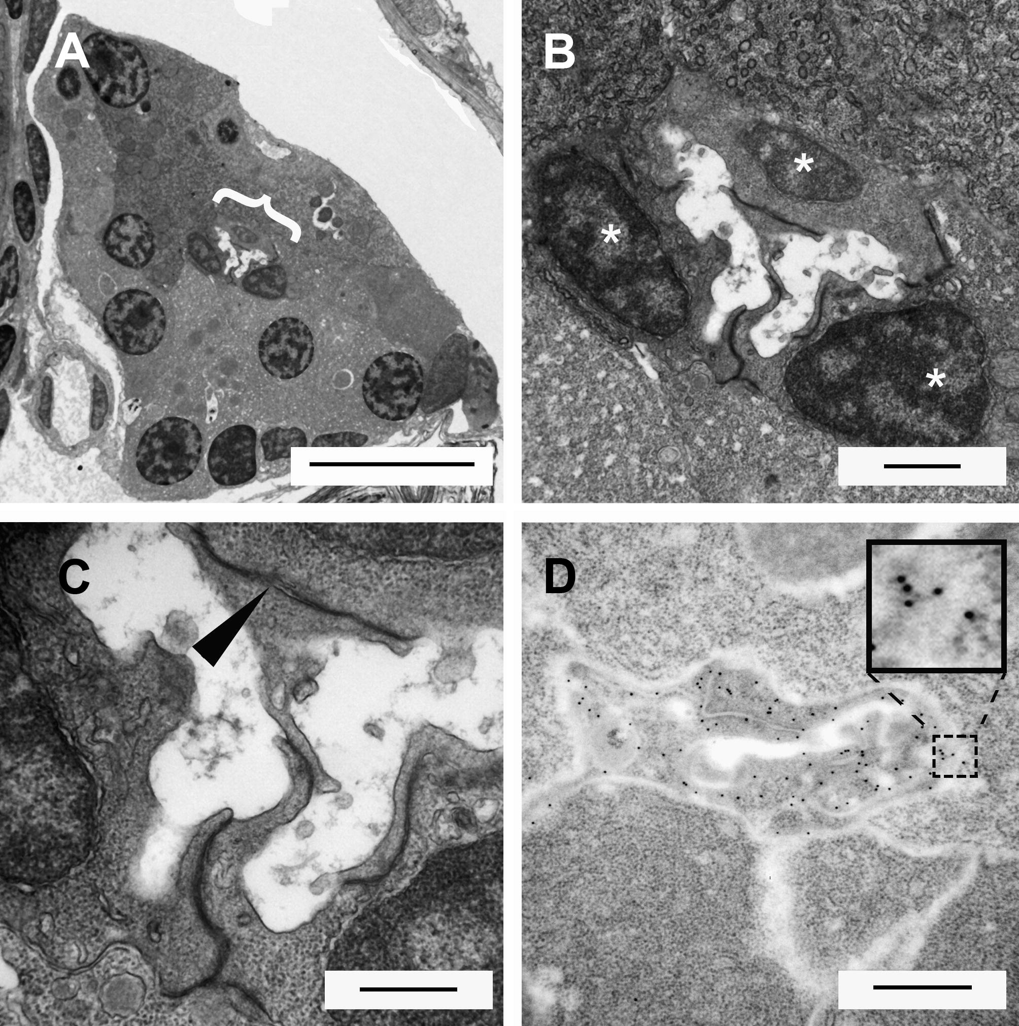
Fig. 5 Electromicrographs of transverse sections through the 10 dpf larval pancreas. (A) At low power the whole transverse section of the pancreas appears triangular. Large nuclei and zymogen granules characterize acinar cells of the exocrine pancreas. Right in the middle of the pancreas the lumen of the pancreatic duct can be seen ({, scale bar = 10 μm). (B) Close up of lumen region in (A). Three cells can be seen around the circumference of the lumen (scale bar = 1 μm). (C) These duct localized cells have projection into the lumen. Wherever the cell membranes of these cells are in direct contact with each other, distinctive tight junctions are observed (►, scale bar = 500 nm). (D) Immunoelectron microscopy using a gold conjugated secondary to detect GFP protein in a pancreas of a Tg(Tp1bglob:eGFP)um14 larva (scale bar = 500 nm). The ultra-structure appears slightly different due to changes in fixation to permit immunolabelling. Gold particles appear very dark and circular (see inset enlargement) and are detected throughout the cells lining the duct. These cells are morphologically similar and in the same position as the cells previously observed by EM. This demonstrates that the previously described PNCs are located in the lining of the pancreatic duct.
Reprinted from Mechanisms of Development, 126(10), Parsons, M.J., Pisharath, H., Yusuff, S., Moore, J.C., Siekmann, A.F., Lawson, N., and Leach, S.D., Notch-responsive cells initiate the secondary transition in larval zebrafish pancreas, 898-912, Copyright (2009) with permission from Elsevier. Full text @ Mech. Dev.