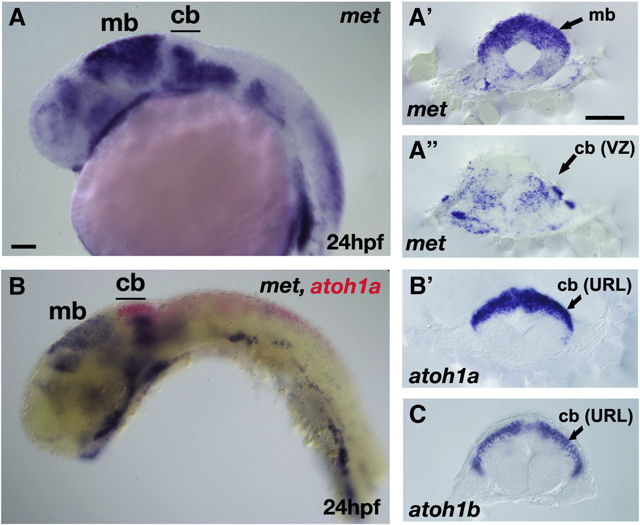
Fig. S1 Expression of the met receptor gene in medial rhombomere 1 (presumptive cerebellum). Lateral view (A and B) and transverse sections (A′, A″, B′ and C) at the level of the midbrain (A′) and cerebellum (A″, B′ and C) of in situ hybridization of 24 hpf embryos showing expression of the met receptor gene in purple (A, A′, A″, B and B′), atoh1a (red in B and purple in B′) and atoh1b (purple in C). In the midbrain region, met expression is abundant in the dorsal domain (A and A′), while in r1, expression is confined to the medial domain (A and A″) corresponding to the ventricular zone of the cerebellum. Double in situ hybridization (B) with met and atoh1a probes shows complementary expression within the cerebellar region, with met being expressed medially in the ventricular zone region and atoh1a being expressed dorsally in the upper rhombic lip region. Single in situ hybridizations with met (A″), atoh1a (B′) and atoh1b (C) also show the complementary expression domains of the met and atoh1 genes. Anterior is to the left and dorsal (transverse sections) is up in all Supplementary figures. mb, midbrain; cb, cerebellum; VZ, ventricular zone; URL, upper rhombic lip. Scale bar, 50 μm.
Reprinted from Developmental Biology, 335(1), Elsen, G.E., Choi, L.Y., Prince, V.E., and Ho, R.K., The autism susceptibility gene met regulates zebrafish cerebellar development and facial motor neuron migration, 78-92, Copyright (2009) with permission from Elsevier. Full text @ Dev. Biol.

