
Fig. S6
- ID
- ZDB-IMAGE-091113-110
- Publication
- Sanek et al., 2009 - Zebrafish zic2a patterns the forebrain through modulation of Hedgehog-activated gene expression
- All Figures
- Figures for Sanek et al., 2009
|
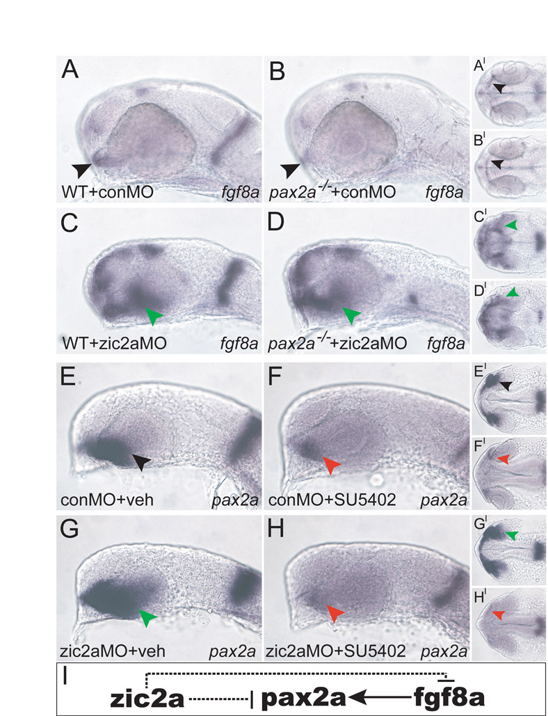
Fig. S6 The epistatic relationships between zic2a, pax2a and fgf8a. (A-H) conMO-injected WT siblings exhibit normal fgf8a expression in the OS and MHB (A, 58/60 embryos, n=2). pax2-/- mutant embryos injected with conMO express fgf8a in the OS, but not in the MHB (B, 20/20 embryos, n=2). WT siblings injected with zic2aMO show expanded fgf8a (C, green arrow, 55/80 embryos, n=2). pax2-/- mutants injected with zic2aMO also have expanded fgf8a in the ventral retina (D, 13/23 embryos, n=2), but lack MHB expression. Vehicle-treated embryos injected with conMO display normal pax2a expression (E, 35/36 embryos, n=4), whereas conMO-injected embryos treated with SU5402 show reduced pax2a (F, 34/37 embryos, n=4). Vehicle-treated zic2aMO-injected embryos have expanded pax2a expression (G, 28/32 embryos, n=4), but zic2aMO-injected and SU5402-treated embryos have reduced pax2a expression (H, 33/36 embryos, n=4). (I) The proposed regulatory relationship between pax2a and fgf8a downstream of zic2a. Arrowheads point to the OS. A-H are lateral views, anterior to the left. A′-H′ are ventral views of the same embryos, anterior to the left. Embryos in A-D are at prim-5 and embryos in E-H are at -prim-1.