
|
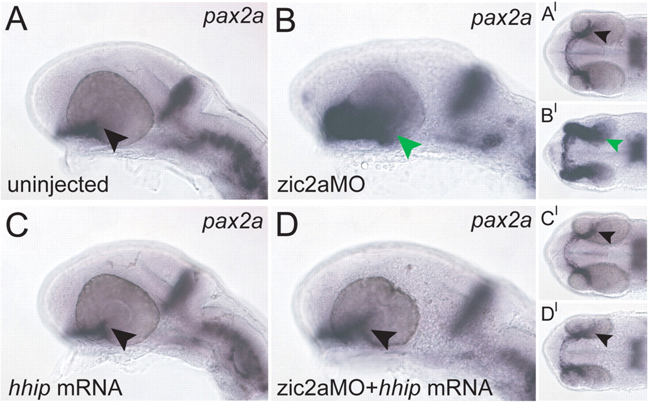
Fig. 10 Exogenous Hh antagonist rescues OS and retinal patterning in Zic2a-depleted embryos. (A) Wild-type pax2a expression. (B-D) Embryos injected with zic2aMO show expanded pax2a (B, 43/43 embryos, n=3), whereas embryos injected with hhip mRNA show normal pax2a expression (C, 8/8 embryos, n=1). Embryos co-injected with zic2aMO and hhip mRNA show normal pax2a expression (D, 30/41 embryos, n=3). A-D are lateral views, anterior to the left. A′-D′ are ventral views of the same embryos, anterior to the left. Arrowheads mark the posterior limit of pax2a expression in the retina. All embryos are at prim-5.