
Fig. S5
- ID
- ZDB-IMAGE-091012-13
- Publication
- Jacoby et al., 2009 - The zebrafish dystrophic mutant softy maintains muscle fibre viability despite basement membrane rupture and muscle detachment
- All Figures
- Figures for Jacoby et al., 2009
|
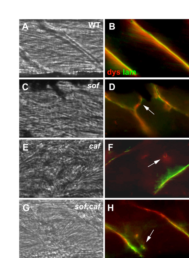
Fig. S5 Analysis of fibre detachment in sof;caf double mutants. (A-H) Antibody co-staining with anti-dystrophin (dys) and anti-laminin (lam) in mutant embryos at 120 hpf. (A,B) Wild-type sibling; (C,D) sof mutant embryo; (E,F) caf mutant embryo; (G,H) sof;caf double mutant embryo. Ectopic fibre termination (EFT)-like structures are seen in sof mutants (arrow in D), whereas detached fibres in caf and sof;caf embryos display only fragmentary basement membrane staining (arrows in F,H). (A,C,E,G) DIC images; (B,D,F,H) merge of fluorescence images. All panels show lateral views with anterior to the left.