Image
Figure Caption
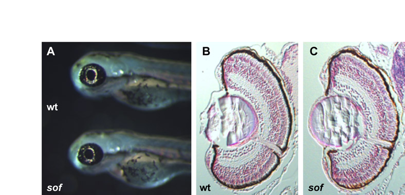
Fig. S2 Histology of the sof mutant eye is normal. (A) Lateral views of the head of wild-type sibling (top) and mutant (bottom) embryos at 72 hpf. (B) Transverse section of a wild-type sibling eye. (C) Transverse section of a sof mutant eye. The mutant eye appears normal, with no disruption of basement membranes or lens malformation, taking into account sectioning artefacts. Sections were stained with Hematoxylin and Eosin.
Acknowledgments
This image is the copyrighted work of the attributed author or publisher, and
ZFIN has permission only to display this image to its users.
Additional permissions should be obtained from the applicable author or publisher of the image.
Full text @ Development

