
|
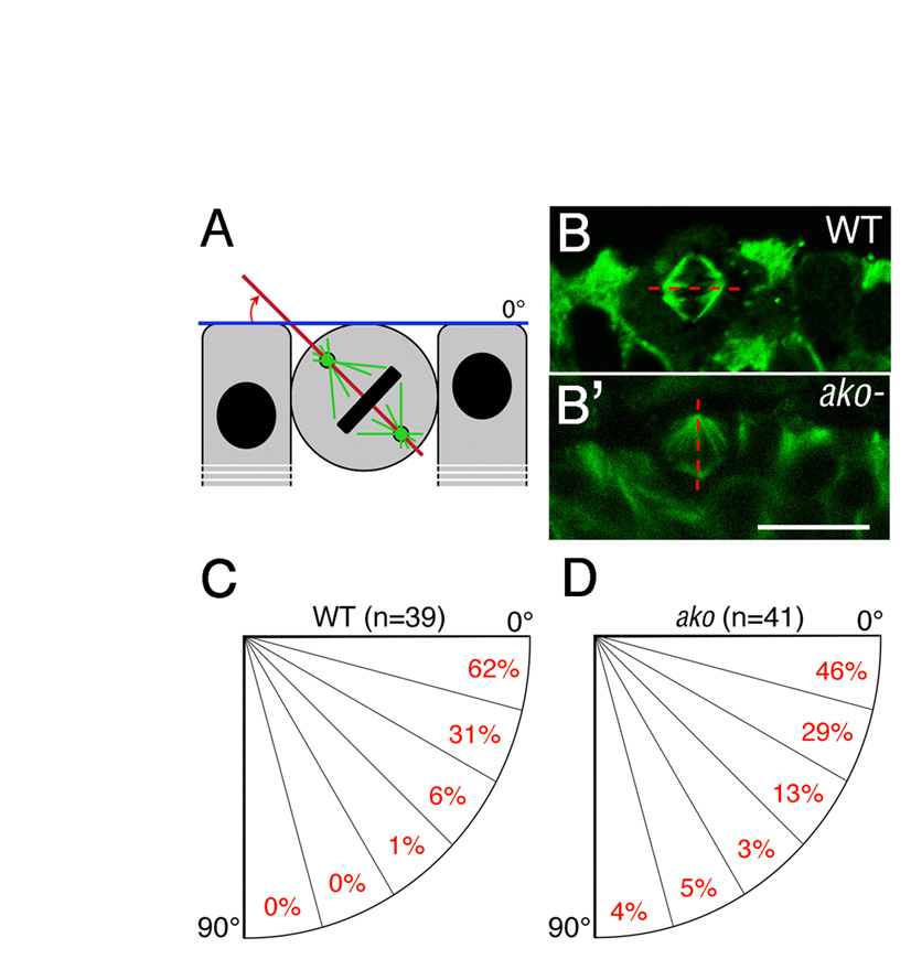
Fig. S4 ale oko function in mitotic spindle orientation. (A) Schematic representation of mitotic spindle orientation relative to the apical surface of the retinal neuroepithelium. The angle between the spindle axis (red line) and the apical surface of the retinal neuroepithelium (blue line) was measured. (B,B′) Transverse sections through wild-type (B) and ako mutant (B′) neuroepithelia were stained with an anti-α-tubulin antibody to visualize mitotic spindles (green). Apical is up. (C,D) Quantification of spindle orientation in wild-type (C) and ako mutant (D) retinae. The alignment of the mitotic spindle is clearly less consistent in akojj50 retinae. Scale bar: 10 μm.