
Fig. S1
- ID
- ZDB-IMAGE-090904-23
- Publication
- Jülich et al., 2009 - Control of extracellular matrix assembly along tissue boundaries via Integrin and Eph/Ephrin signaling
- All Figures
- Figures for Jülich et al., 2009
|
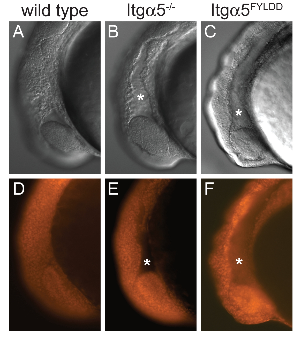
Fig. S1 Impairment of itga5 function leads to a phenotype in the zebrafish head. Lateral views of 9-somite stage zebrafish embryos. DIC (A-C) and widefield (D-F) images of cell nuclei labeled with propidium iodide. (A,D) Wild type. (B,E) In itga5-/- embryos, there is a deficit of cells posterior to the eye (asterisks). This phenotype is evident in mutant embryos with virtually unrecognizable somite phenotypes (presumably due to maternal rescue and/or genetic background). Thus, head development is more sensitive than somitogenesis to the loss of itga5. (C,F) This head phenotype is observed in wild-type embryos that express itga5FYLDD, demonstrating the antimorphic activity of this integrin variant.