
IMAGE
Fig. S3
- ID
- ZDB-IMAGE-090817-14
- Publication
- McMahon et al., 2009 - Lmx1b is essential for survival of periocular mesenchymal cells and influences Fgf-mediated retinal patterning in zebrafish
- All Figures
- Figures for McMahon et al., 2009
Image
|
Figure Caption
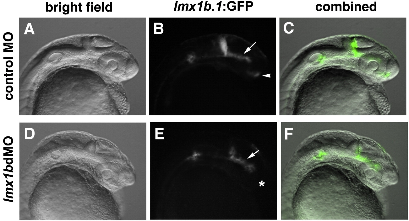
Fig. S3 lmx1b.1:GFP fluorescence is lost in the optic stalk region, but maintained in the mid-ventral diencephalon of lmx1bdMO embryos. (A, D) Bright field images of control or lmx1bdMO embryos for the Tg(- 5 kb lmx1b.1:GFP)mw13 line. (B, E) GFP-fluorescent images of the same embryos. (C, F) Combined bright field and fluorescent images. Arrow indicates the mid-ventral diencephalon, and arrowhead indicates optic stalk region. Asterisk indicates lack of GFP expression in the optic stalk region of lmx1bdMO embryos.
Acknowledgments
This image is the copyrighted work of the attributed author or publisher, and
ZFIN has permission only to display this image to its users.
Additional permissions should be obtained from the applicable author or publisher of the image.
Reprinted from Developmental Biology, 332(2), McMahon, C., Gestri, G., Wilson, S.W., and Link, B.A., Lmx1b is essential for survival of periocular mesenchymal cells and influences Fgf-mediated retinal patterning in zebrafish, 287-298, Copyright (2009) with permission from Elsevier. Full text @ Dev. Biol.