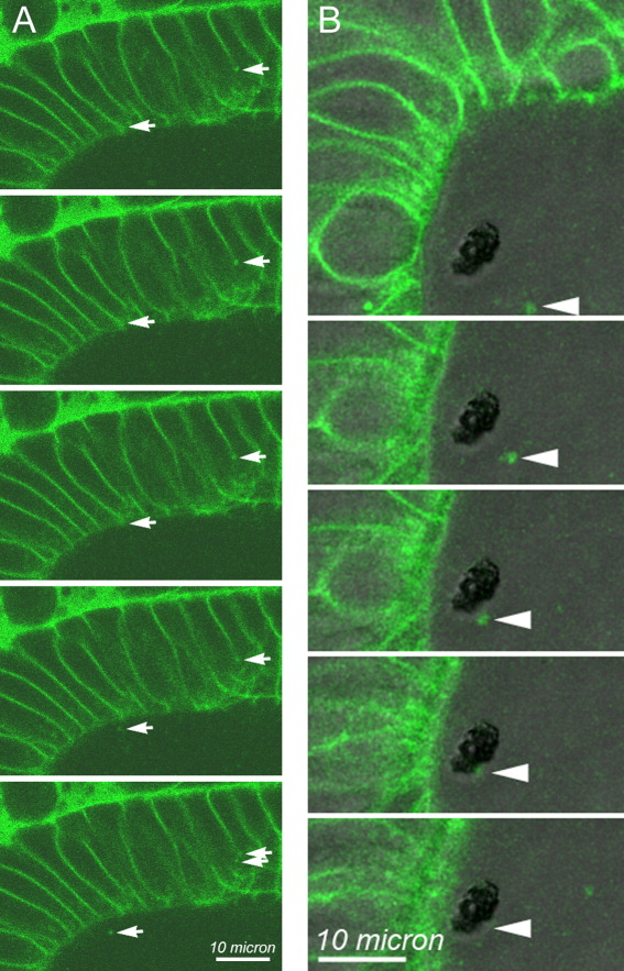
Fig. 6 Time lapse imaging of BODIPY-ceramide-labeled vesicular structures suspended within the otolymph and fusing with the otolith. Using the same time-lapse images used to determine size and numbers of vesicular structures within the otolymph of 22-hpf control embryos, movement of individual BODIPY-ceramide-labeled structures was examined. BODIPY-labeled vesicles were seen moving within otic epithelial cells, being exported from otic epithelial apical cell surfaces into the otolymph, moving within the otolymph, and fusing with the growing otolith. Examples of these events are shown in time-lapse sequence. DIC and BODIPY fluorescence images were collected every 2 sec. The first image of each time sequence is shown at the top and proceeds every 2 sec in sequential images from top to bottom. A: Only the BODIPY fluorescence images are shown for this sequence. Arrows on the left side of each panel indicate a BODIPY-ceramide-labeled structure at the lateral cell surface of an otic epithelial cell that moves in a generally apical direction. Arrows on the right side of each panel indicate the apico-lateral region of the cell that appears to generate a BODIPY-labeled punctate vesicle-like structure that enters the otolymph in the final two frames of this sequence. B: DIC and BODIPY fluorescence images were combined into a single image shown in each frame. In B, the dark, unstained structure in the otolymph is the otolith. Single image planes are presented, and the specific image plane within the volume varied over the time sequence to optimize the visualization of the particular BODIPY-labeled puncta highlighted by the arrowheads in each panel. All other image planes were examined to insure that the indicated vesicular structure was not merely moving out of the plane of focus, but appeared to adhere and fuse at the surface of the otolith. Scale bar = 10 μm.
Image
Figure Caption
Acknowledgments
This image is the copyrighted work of the attributed author or publisher, and
ZFIN has permission only to display this image to its users.
Additional permissions should be obtained from the applicable author or publisher of the image.
Full text @ Dev. Dyn.

