
Fig. 8
- ID
- ZDB-IMAGE-090804-65
- Publication
- Ng et al., 2009 - Zebrafish mutations in gart and paics identify crucial roles for de novo purine synthesis in vertebrate pigmentation and ocular development
- All Figures
- Figures for Ng et al., 2009
|
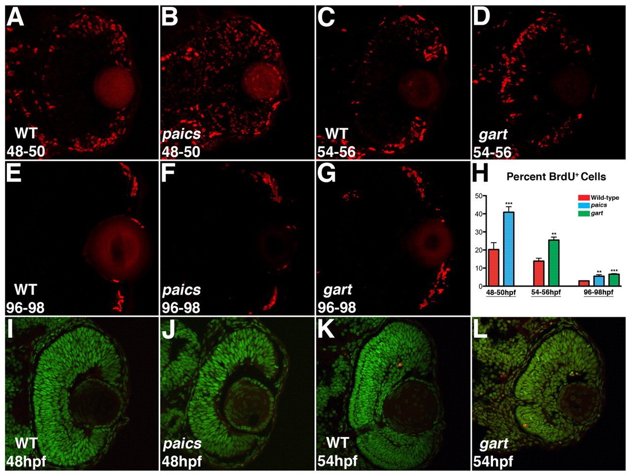
Fig. 8 Cell proliferation and apoptosis in gart and paics mutants. (A,C) BrdU exposures 48-50 hpf and 54-56 hpf indicate that in the wild-type eye most proliferative cells are located at the retinal periphery. (B) In paics and (D) gart mutants, BrdU+ cells are located throughout the central retina. (E-G) BrdU exposures 96-98 hpf localized proliferative cells in the wild-type and both paics and gart mutant retinas to the retinal periphery. (H) Quantifying the percentage of BrdU+ cells in each retina identified significant increases in the paics and gart mutants at each time point (** P<0.05, *** P<0.001). (I-L) TUNEL labeling to identify apoptotic nuclei. No increases in apoptosis over wild-type levels were observed in paics mutants at 48 hpf (I,J) or gart mutants at 54 hpf (K,L).