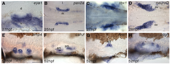
Fig. S1 Early patterning of the otic vesicle appears normal in lte mutants. Whole-mount in situ hybridisation shows that the expression of early patterning markers in the otic vesicle is indistinguishable between siblings and mutants (n=30 plus embryos per batch from a cross between heterozygous parents; staining was identical in all embryos within a batch). Normal expression is seen for eya1 ventrally (A), pax2a medially (B) and tbx1 laterally (C) at 25-27 hpf. The statoacoustic ganglion delaminates as usual from the otic vesicle, marked by neurod expression at 28 hpf (D). bmp4 and cahz mark the cristae (E, white asterisks) and maculae (F), respectively, at 52 hpf. The endolymphatic duct, marked by expression of foxi1 (G), appears to evaginate normally. The semicircular canal projections also form normally (H, black asterisks). d, dorsal; l, lateral; m, medial; v, ventral; am, anterior macula; ed, endolymphatic duct; ov, otic vesicle; pm, posterior macula. A,E-H, lateral views; B-D, ventral views. Scale bars: 50 μm.
Image
Figure Caption
Acknowledgments
This image is the copyrighted work of the attributed author or publisher, and
ZFIN has permission only to display this image to its users.
Additional permissions should be obtained from the applicable author or publisher of the image.
Full text @ Development

