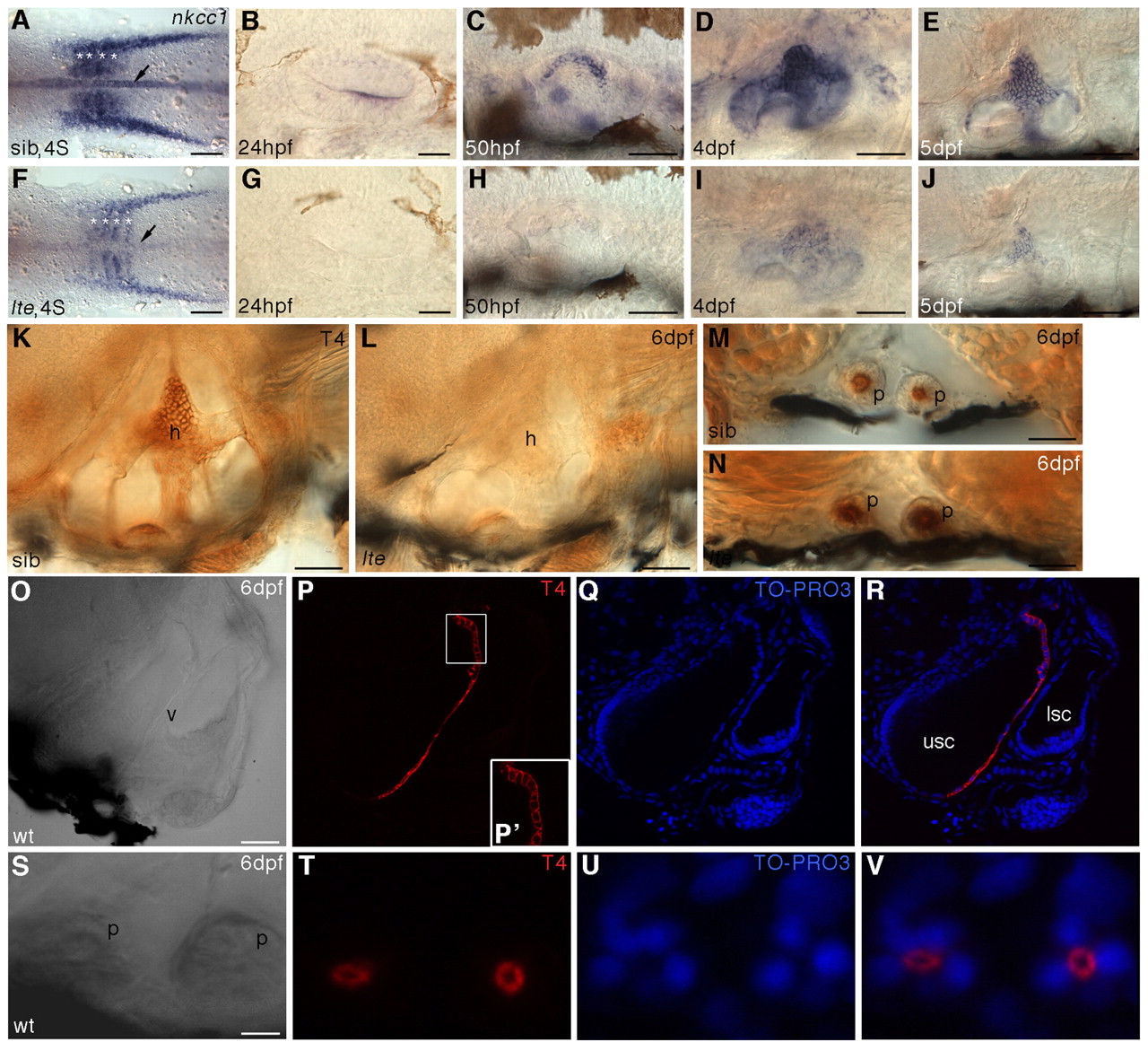
Fig. 4 Expression of nkcc1 transcript and protein immunoreactivity are lost in lte mutants. (A-J) Whole-mount in situ hybridisation for nkcc1 reveals a reduction or loss of expression in lte mutants at all stages examined. (A,F) Four-somite stage (4S). Expression is reduced in the notochord (arrows) and somites (asterisks) in putative mutants. (B-E,G-J) Expression is reduced or lacking in the lte mutant ear from 24 hpf to 5 dpf. (K-V) Immunostaining with the T4 (Nkcc1/2) antibody. Immunoreactivity is lost at 6 dpf in the lte mutant ear (K,L). Expression of Nkcc2 in the pronephric ducts is unaffected in mutants (transverse sections M,N). (O-R) Confocal transverse section through the wild-type ear at 6 dpf; O is a brightfield image and R is an overlay of P and Q. Nkcc1 expression is restricted to the cells lining the medial face of the ventral pillar (P,R, red). TO-PRO-3 staining of nuclei (Q,R, blue) confirms that expression is restricted to the cell membrane and is highest basolaterally (P′). (S-V) Confocal transverse section through the pronephric ducts at 6 dpf showing that Nkcc2 expression is restricted to the apical cell membrane. h, semicircular canal hub; p, pronephric duct; v, ventral pillar; usc, utriculosaccular chamber; lsc, lateral semicircular canal lumen. Scale bars: 200 μm in A,F; 50 μm in B-E,G-L,O-R; 25 μm in M,N; 12.5 μm in S-V.
Image
Figure Caption
Figure Data
Acknowledgments
This image is the copyrighted work of the attributed author or publisher, and
ZFIN has permission only to display this image to its users.
Additional permissions should be obtained from the applicable author or publisher of the image.
Full text @ Development

