
Fig. 2
- ID
- ZDB-IMAGE-090804-20
- Publication
- Abbas et al., 2009 - Nkcc1 (Slc12a2) is required for the regulation of endolymph volume in the otic vesicle and swim bladder volume in the zebrafish larva
- All Figures
- Figures for Abbas et al., 2009
|
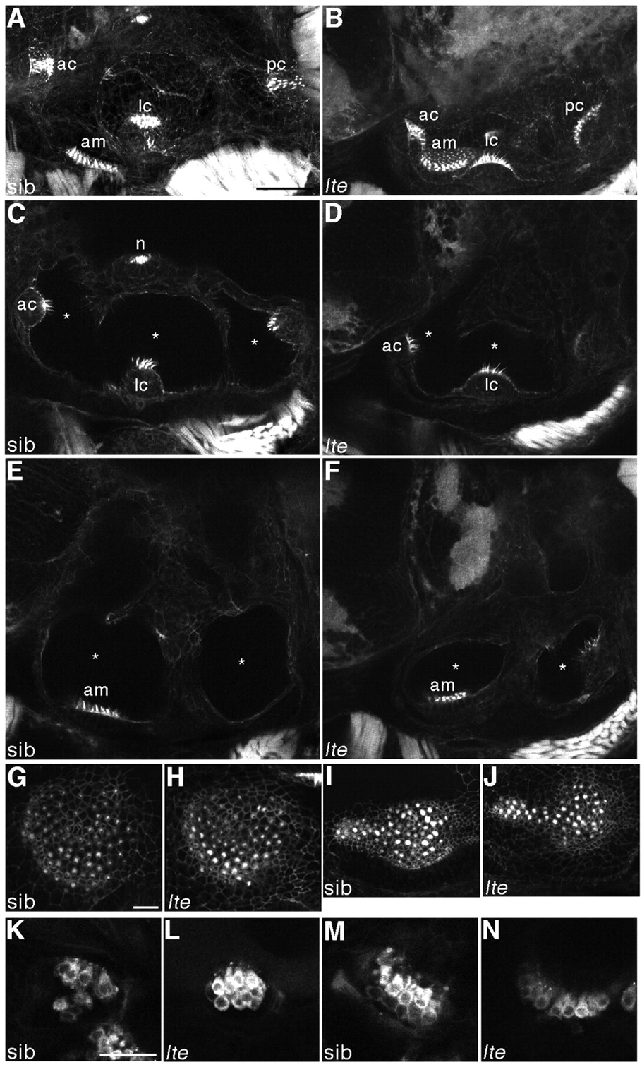
Fig. 2 Hair cells do not degenerate and are still functional in lte mutant ears. (A-J) Staining with FITC-phalloidin labels hair cells at 6 dpf. In both siblings (A,C,E,G,I) and lte mutants (lteto27d in B,D,F; ltetg414b in H,J), actin-rich hair bundles are visible in all five sensory patches. Enlargements of the anterior (G,H) and posterior (I,J) maculae show equivalent numbers of differentiated hair cells in siblings and lte mutants. Asterisks (C-F) mark the reduction in the volume of the otic vesicle lumina in the mutant. (K-N) Live staining with FM1-43 marks hair cell endocytosis at 5 dpf, labelling functional cells in the cristae (K,L) and the anterior macula (M,N) in siblings (K,M) and mutants (L,N). A,B,G-J are projections of confocal z-stacks; C-F,K-N are individual optical sections, in which not all sensory patches are visible. am, anterior macula; ac, anterior crista; lc, lateral crista; pc, posterior crista; n, neuromast. Scale bars: 50 μm in A-F; 25 μm in G-N.