
Fig. 1
- ID
- ZDB-IMAGE-090804-19
- Publication
- Abbas et al., 2009 - Nkcc1 (Slc12a2) is required for the regulation of endolymph volume in the otic vesicle and swim bladder volume in the zebrafish larva
- All Figures
- Figures for Abbas et al., 2009
|
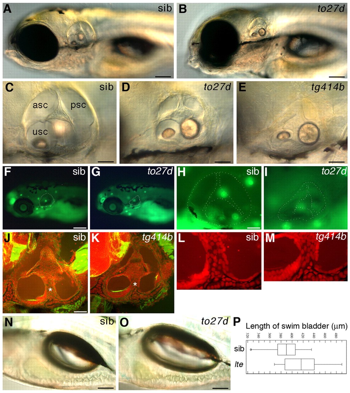
Fig. 1 lte mutant zebrafish larvae have a collapsed ear at 5 dpf and an increased swim bladder volume. (A-D) Live images of lteto27d homozygous mutant (B,D) and phenotypically wild-type sibling (sib) (A,C) embryos at 5 dpf. (E) Ear collapse in the tg414b allele at 5 dpf is slightly more severe than in to27d. (F-I) Acridine Orange staining (green) indicates that there is no increase in cell death in the ear (white dashed outline) in lte mutants as compared with siblings at 5 dpf (F,H compared with G,I). Background staining in live neuromasts can be seen in both genotypes (green spots). (J-M) FITC-phalloidin (green)/propidium iodide (red) staining reveals similar architecture and number of cells within the otic epithelium of tg414b and siblings at 5 dpf. (L,M) Enlargement of the ventral pillar (white asterisks in J,K). (N,O) Live images showing the increase in swim bladder volume in lteto27d mutants. (P) The average swim bladder length increases from 390 μm in sibling embryos to 418 μm in the mutants (n=12 siblings, n=12 mutants; P=0.03, Student′s t-test). Horizontal lines indicate the distribution of swim bladder lengths; vertical lines indicate the population mean; boxes indicate standard deviation. asc, anterior semicircular canal; psc, posterior semicircular canal; usc, utriculosaccular chamber. Scale bars: 125 μm in A,B; 50 μm in C-E,H-K; 200 μm in F,G; 100 μm in N,O.