
IMAGE
Fig. S4
- ID
- ZDB-IMAGE-090804-11
- Publication
- Kurrasch et al., 2009 - Neuroendocrine transcriptional programs adapt dynamically to the supply and demand for neuropeptides as revealed in NSF mutant zebrafish
- All Figures
- Figures for Kurrasch et al., 2009
Image
|
Figure Caption
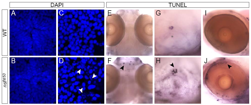
Fig. S4 Cell death is observed in sensory neurons in nsf mutant larvae. DAPI stain in the hypothalamus (A, B) and the posterior tuberculum of wild type and nsfst53 sectioned (12 μm horizontal sections) 5dpf zebrafish is shown. Morphological changes associated with cell death, such as cell asymmetry and chromatin condensation is noted (white arrowheads; D). Cell death is not observed in the hypothalamus (A,B), but is observed in olfactory terminals (F), otic capsule (H) and photoreceptor cells (J) of mutant zebrafish. Images representative of duplicate experiments, n≥5 per genotype.
Acknowledgments
This image is the copyrighted work of the attributed author or publisher, and
ZFIN has permission only to display this image to its users.
Additional permissions should be obtained from the applicable author or publisher of the image.
Full text @ Neural Dev.