
Image
|
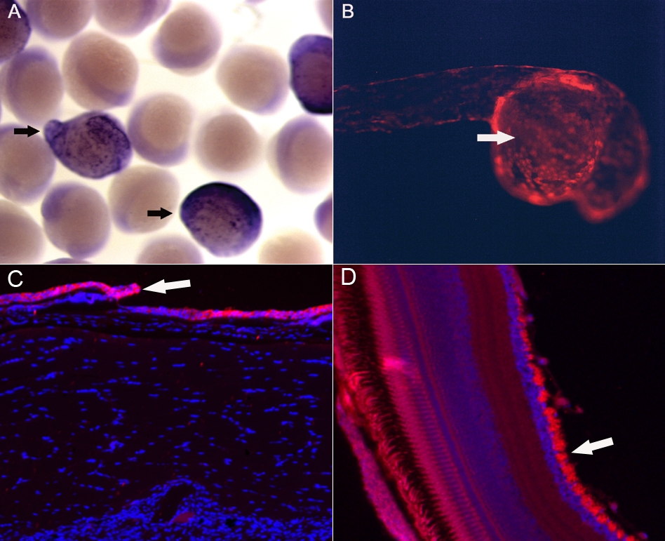
Figure Caption
Fig. S3 Expression patterns of transgenic line A. The data indicated that the cytokeratin 4 promoter drove epithelial cells-specific expression (arrows) of smoa1-EGFP as shown by in situ hybridization against EGFP in 12 hpf F1 embryos (A), and of tdTomato in a 24 hpf embryo generated by crossing the Tg(krt4:Gal4VP16;14 x UAS:smoa1-EGFP) and Tg(UAS:tdTomato) transgenic fish (B). At adult stage, GFP was detected predominantly in skin epithelial cells (C, arrow) and the retinal ganglion cells (D, arrow).
Acknowledgments
This image is the copyrighted work of the attributed author or publisher, and
ZFIN has permission only to display this image to its users.
Additional permissions should be obtained from the applicable author or publisher of the image.
Open Access.
Full text @ Mol. Cancer