
|
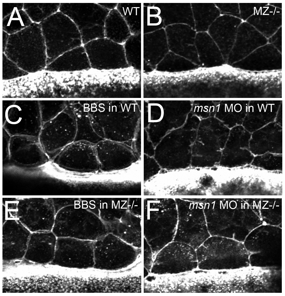
Fig. S3 In contrast to loss of msn1 or treatment with blebbistatin, loss of epcam does not compromise the constriction of marginal EVL cells. (A–F) Fluorescent confocal images of phalloidin stained maternal zygotic-EpCAM mutant (MZ-/-) and wild type (WT) embryos at 70% epiboly. The leading EVL cells of both, mutant and WT embryos are constricted by an actin-myosin string as indicated by the presence of cells with shorter leading than trailing sides (A,B). This is in contrary to the release of constriction caused by in wild-type embryos upon treatment with 15 μg/ml blebbistatin (C) or injection of msn1 MO (D) [10], suggesting that the EVL epiboly defects of MZepcam and msn1 mutants have a different cellular basis, and that the phenotype of MZepcam mutants is not caused by reduced actin-myosin constriction at the EVL-YSL interface. Consistent with this notion, injection of msn1 MO or blebbistatin treatment in MZepcam mutants, while releasing marginal constrictions (E,F), failed to synergistically enhance the EVL epiboly defects, but rather had pure additive effects (data not shown). Another possibility would have been that the epiboly defects of MZepcam mutants are due to increased/precocious, rather than reduced actin-myosin constriction at the EVL-YSL interface. However, applying increasingly lower amounts of msn1 MO or blebbistatin to MZepcam mutants, we never obtained an alleviation of the EVL epiboly defects (judged by the extrusion of the vegetal-most part of the yolk at late gastrula stages; compare with Figure 5B), also making this possibility very unlikely.