
|
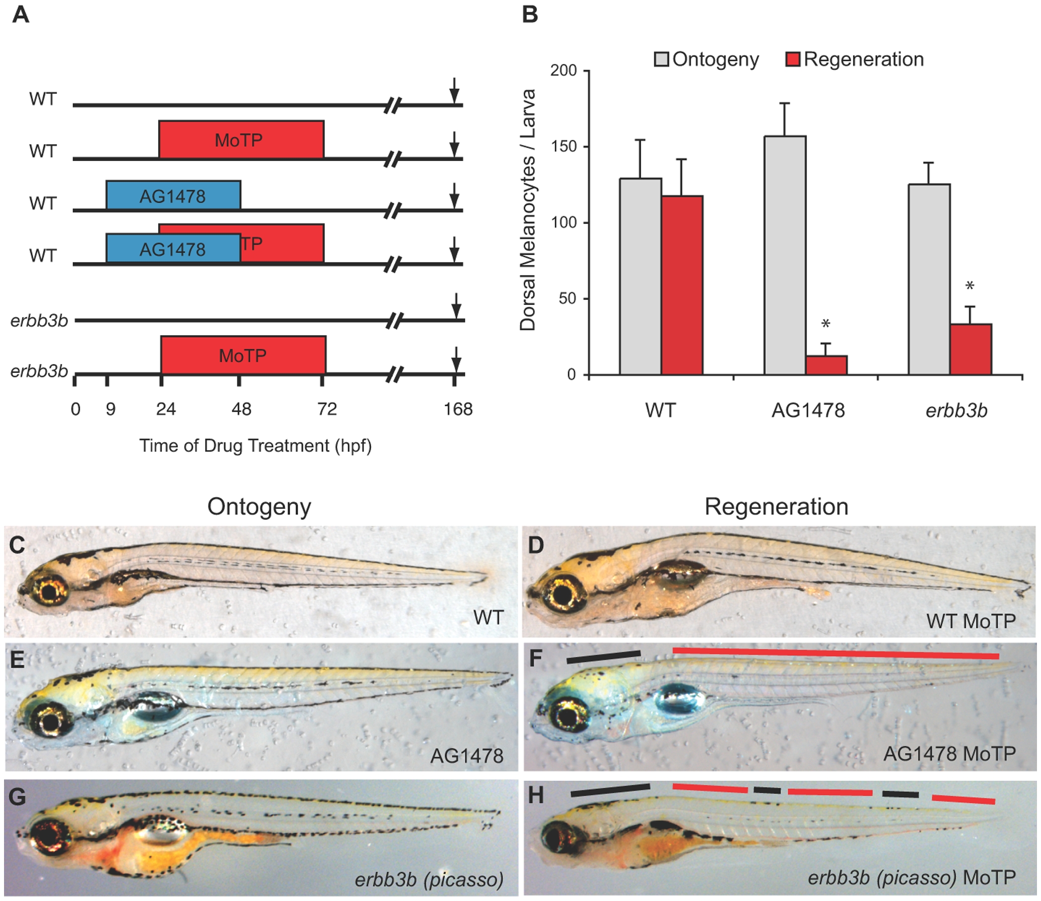
Fig. 1 ErbB signaling is required for larval melanocyte regeneration.
(A) Cartoon of drug treatment timeline for assaying melanocyte ontogeny and regeneration. Arrow indicates when embryos were collected for photos and melanocyte counts, in this case at 168 hpf. (B) Quantitation of average dorsal melanocytes from somites 1–26 for each treatment in (A) for melanocyte ontogeny (gray) and melanocyte regeneration (red). Error bars represent standard deviation, * represents P<0.05 (Student t-test, N = 10). Photos of representative larvae at 168 hpf for ontogeny (C, E, G) and regeneration (D, F, H). WT larvae regenerate nearly completely (compare D to C). WT treated with AG1478 (E, F) and erbb3b mutants (G, H) largely fail to regenerate but have normal ontogenetic number of melanocytes. A few regeneration melanocytes are observed in the head and sporadically in parts of the trunk (black bars in F and H) but are mostly absent throughout the trunk (red bars in F and H).