
IMAGE
Fig. S2
- ID
- ZDB-IMAGE-090717-20
- Publication
- Lamont et al., 2009 - Antagonistic interactions among Plexins regulate the timing of intersegmental vessel formation
- All Figures
- Figures for Lamont et al., 2009
Image
|
Figure Caption
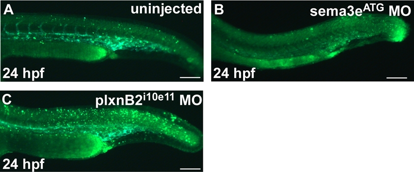
Fig. S2
Assessment of non-specific cell death in sema3ee3i3 and plxnB2i10e11 morphant by acridine orange staining.
(A-C) Acridine orange staining of Tg(kdr:GFP)la116 uninjected (A), sema3eATG morphant (B), and plxnB2i10e11 morphant (C) embryos at 24 hpf. Green dots represent cells undergoing apoptosis.
Acknowledgments
This image is the copyrighted work of the attributed author or publisher, and
ZFIN has permission only to display this image to its users.
Additional permissions should be obtained from the applicable author or publisher of the image.
Reprinted from Developmental Biology, 331(2), Lamont, R.E., Lamont, E.J., and Childs, S.J., Antagonistic interactions among Plexins regulate the timing of intersegmental vessel formation, 199-209, Copyright (2009) with permission from Elsevier. Full text @ Dev. Biol.