
Fig. 4
- ID
- ZDB-IMAGE-090710-54
- Publication
- Yabe et al., 2009 - The maternal-effect gene cellular island encodes aurora B kinase and is essential for furrow formation in the early zebrafish embryo
- All Figures
- Figures for Yabe et al., 2009
|
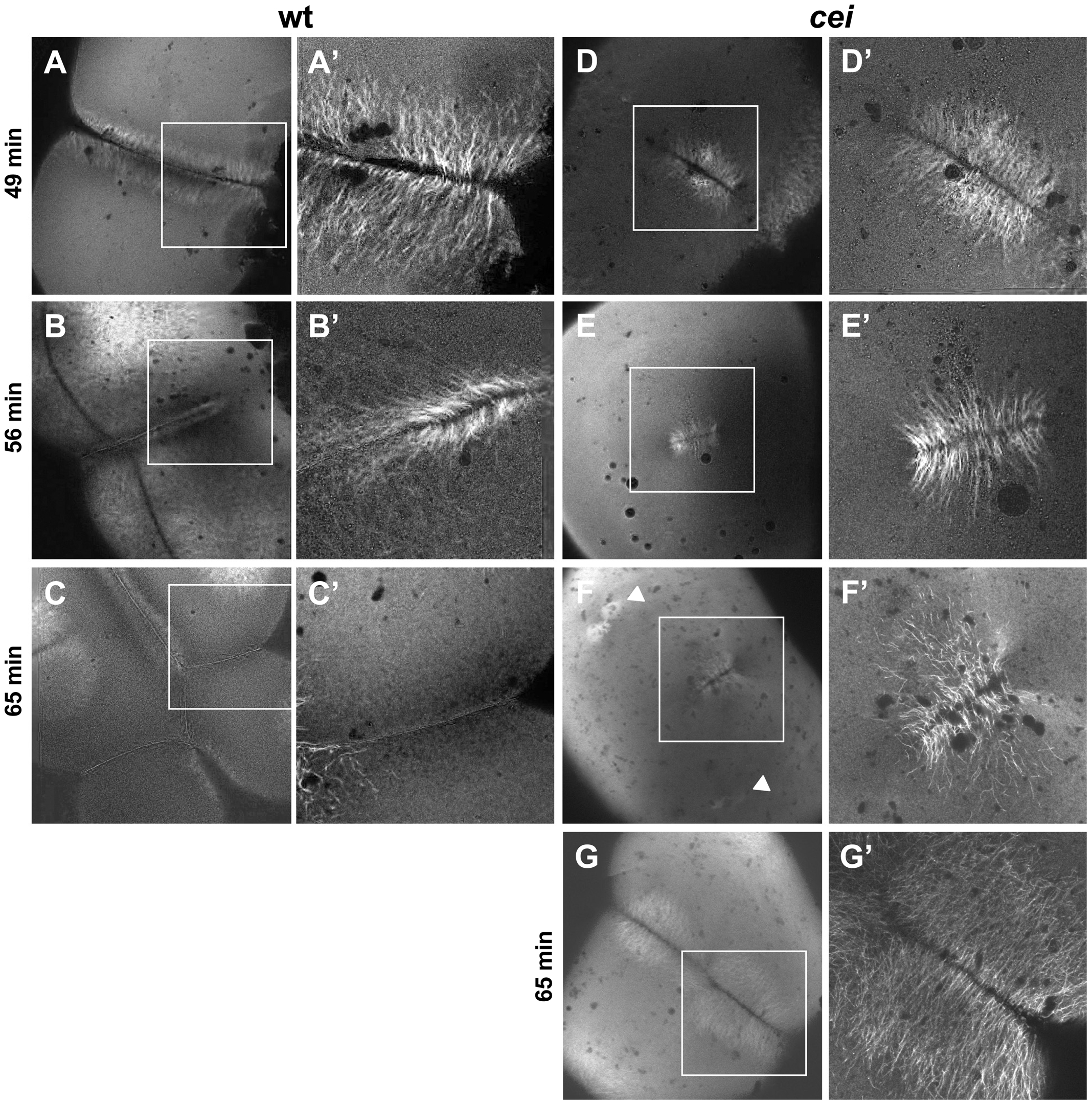
Fig. 4 Defective cytoskeletal dynamics in cei/aurB mutant embryos.
(A–G) High magnification images of wild-type (A–C) and cei mutant (D–G) embryos fixed at the indicated time points and labeled with an anti-α-tubulin antibody. Images show the FMA structure, which after formation of the furrow (A) becomes enriched in the distal region of the wild-type embryo (B) and eventually disassembles (C). In maternal cei mutants, the truncated FMA forms in the center of the blastodisc (D) and neither translocates distally nor becomes disassembled (E,F). Arrowheads in (F) indicate FMA remnants corresponding to the second cleavage planes. A rare maternal cei mutant embryo with a weaker phenotype (G) showing that defects in FMA reorganization and disassembly can be observed even when the furrow encompasses the entirety of the blastodisc.