
IMAGE
Fig. 2
- ID
- ZDB-IMAGE-090710-52
- Publication
- Yabe et al., 2009 - The maternal-effect gene cellular island encodes aurora B kinase and is essential for furrow formation in the early zebrafish embryo
- All Figures
- Figures for Yabe et al., 2009
Image
|
Figure Caption
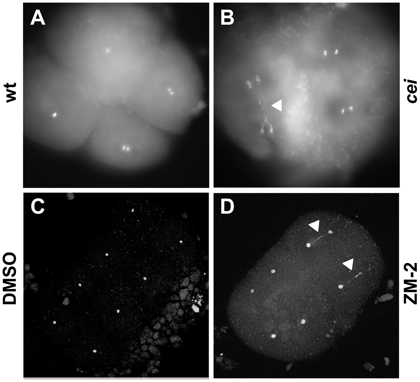
Fig. 2 DNA segregation defects in embryos with reduced cei/aurB function.
Animal views of fixed embryos labeled with DAPI. (A–B) Wild-type (A) and maternally mutant cei (B) embryos at 60 min p.f. (C–D) Solvent- (C) and ZM2- (D) treated embryos at 75 min p.f. Arrowheads indicate DNA bridges.
Figure Data
Acknowledgments
This image is the copyrighted work of the attributed author or publisher, and
ZFIN has permission only to display this image to its users.
Additional permissions should be obtained from the applicable author or publisher of the image.
Full text @ PLoS Genet.