
Fig. S2
- ID
- ZDB-IMAGE-090710-47
- Publication
- Lightcap et al., 2009 - Interaction with LC8 is required for Pak1 nuclear import and is indispensable for zebrafish development
- All Figures
- Figures for Lightcap et al., 2009
|
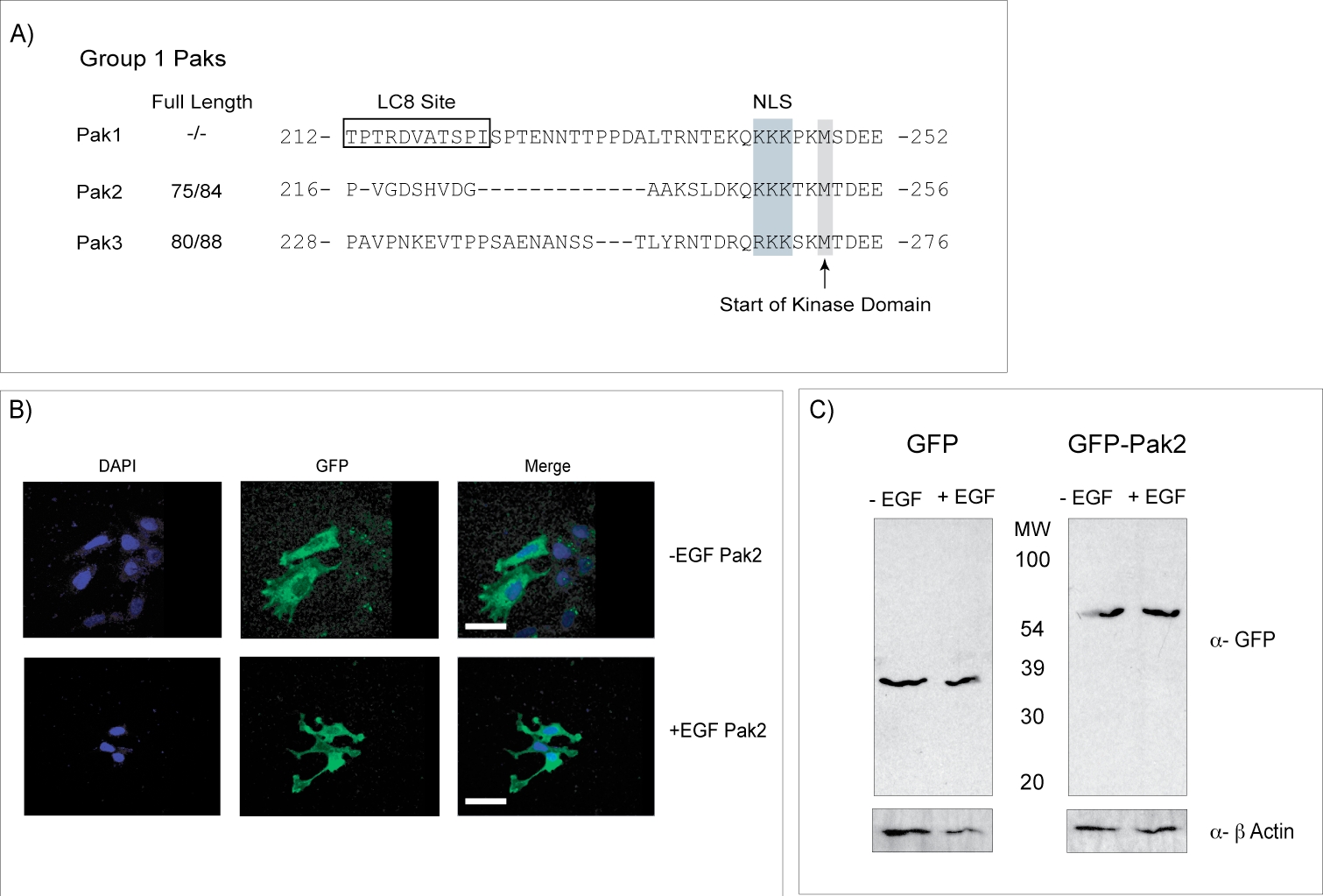
Fig. S2 Pak2 is not cleaved after EGF Stimulation. (A) Sequence alignment of Group1 Pak kinases, highlighting that the Pak1 LC8 binding site (boxed) is absent in Pak2 and Pak3. By contrast, Pak1 and Pak2 share identical nuclear localization sequences (NLS) positioned at the same location upstream of the kinase domain. (B) Representative examples of subcellular distribution of Pak2 as determined by confocal microscopy. Pak2 does not translocate to the nucleus after EGF stimulation in MCF7 cells. (C) Western blot of MCF7 cells expressing either GFP alone or GFP-Pak2 before and after stimulation with EGF. Results show GFP runs at the same molecular weight after EGF treatment, showing that Pak2 is not cleaved in these experiments.