
|
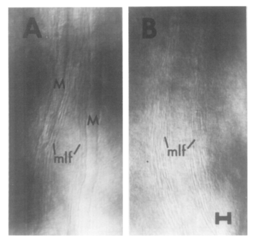
Fig. 1 Nomarski interference contrast views of the hindbrains of 1-week-old irradiated zebra fish larvae. The animals were irradiated at 6 hr after fertilization using a cesium source in a Model M Gammator (Radiation International). The nominal dose rate was 980 rad/min, and the embryos were exposed for 1.0 min. After continued development the animals were examined, as living larvae, by microscopy (Kimmel, 1972). (A) Animal shown with the normal pair of M-axons (M) which accompany the medial longitudinal fascicle (mlf) of finer axons through the hindbrain. (B) Both M-axons are absent, and only the mlf is present. In scoring for missing M-cells, the presence of this fiber pathway was always ascertained to provide an internal control for visibility. Dimension marker = 10 μm.
Reprinted from Developmental Biology, 62(2), Kimmel, C.B., Sessions, S.K., and Kimmel, R.J., Radiosensitivity and time of origin of Mauthner neuron in the zebra fish, 526-529, Copyright (1978) with permission from Elsevier. Full text @ Dev. Biol.