
|
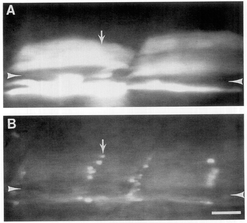
Fig. 1 Wild type muscle fibers cluster receptors when contacted by nic1 motoneurons. (A) Fluorescence illumination showing fluorescein-dextran-labeled donor wild-type muscle fibers (arrow) in a live, unlabeled nic1 host myotome. The remainder of the field of view contains unlabeled nic1 muscle fibers (two are marked with arrowheads). (B) View of same myotome showing R-BTX labeling. R-BTX-labeled AChRs (arrow) are clustered specifically on the wild-type fibers. Arrowheads indicate nic1 muscle fibers (also shown by arrowheads in A) which lack R-BTX labeling of AChRs although they are presumably innervated by the motoneuron contacting adjacent fibers. We made similar observations on 21 single muscle cells and three multiple cell patches in three embryos. In every case, the wild-type fiber developed clusters of AChRs and in no case could we assign a cluster to a mutant fiber. Trunk segment; 80 h; scale bar, 20 μm.
Reprinted from Developmental Biology, 161, Sepich, D.S., Ho, R.K., and Westerfield, M., Autonomous expression of the nic1 acetylcholine receptor mutation in zebrafish muscle cells, 84-90, Copyright (1994) with permission from Elsevier. Full text @ Dev. Biol.