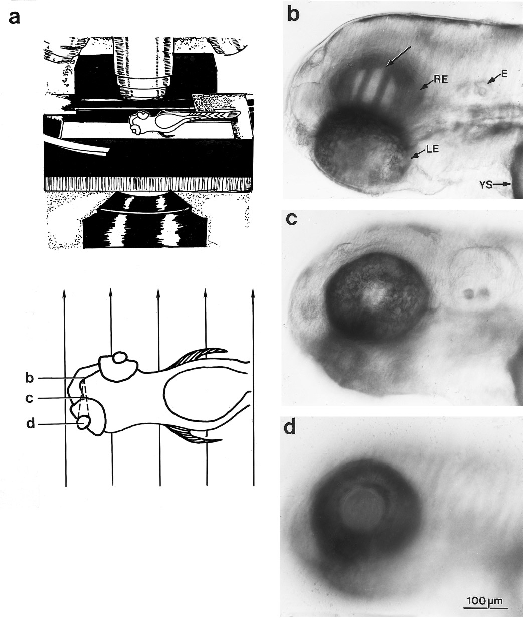
Fig. 7 Image at an inappropriate plane. (a) The upper sketch shows the setup; a larval zebrafish was positioned obliquely on a microscope slide, inside a water-filled chamber (not shown), with one eye oriented downward toward the condenser lens. The lower sketch shows the larva in more detail, with the parallel rays from the SCAD below indicated by the vertical arrows. The image of the SCAD is formed by the ocular lens at the location given by the convergence of the dashed lines. The letters b, c, and d indicate the planes at which the pictures in b, c, and d were focused (d, the lens center; c, the photoreceptor/pigmented epithelium interface; and b, the plane of the image of the grating). (b–d) These are photomicrographs of the same 68 hpf larva, anterior to the left, viewed dorsolaterally with the right eye (RE, toward the top) facing down and the left eye (LE) facing up. The yolk sac (YS) and ear (E) are indicated. The pictures were taken at the three different focal planes shown in a. The image (unlabeled arrow) lay behind the right retina, about twice as far from the lens center as the photoreceptor/pigmented epithelium interface, so the eye was hyperopic (far-sighted). The scale bar applies to b, c, and d.
Reprinted from Developmental Biology, 180, Easter, S.S., Jr. and Nicola, G.N., The development of vision in the zebrafish (Danio rerio), 646-663, Copyright (1996) with permission from Elsevier. Full text @ Dev. Biol.

