
Fig. 2
- ID
- ZDB-IMAGE-090515-11
- Genes
- Publication
- Taneja et al., 1996 - The expression pattern of the mouse receptor tyrosine kinase gene MDK1 is conserved through evolution and requires Hoxa-2 for rhombomere-specific expression in mouse embryos
- All Figures
- Figures for Taneja et al., 1996
|
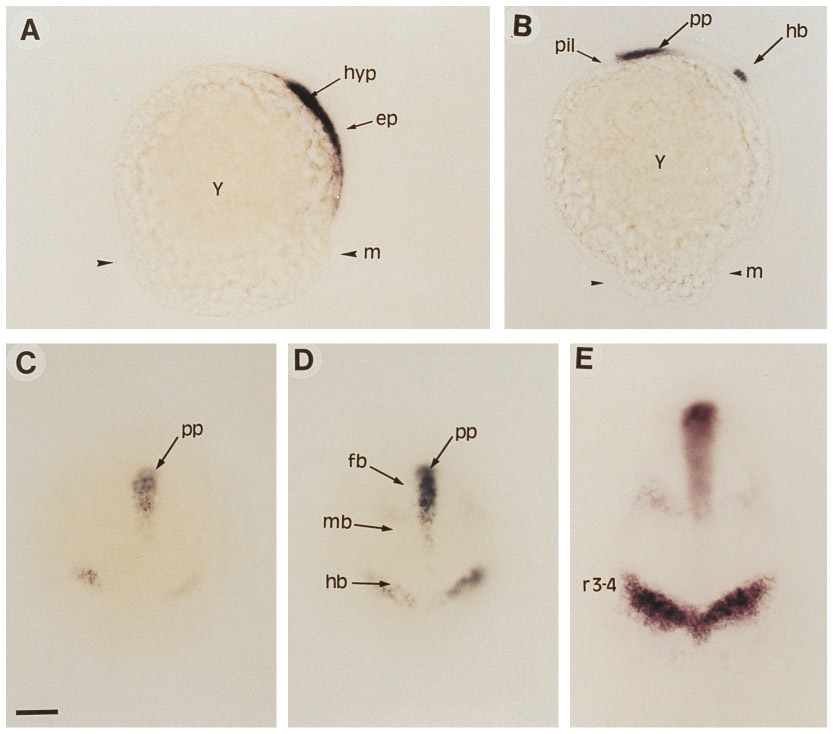
Fig. 2 Early expression pattern of ZDK1 during gastrulation. (A) At 60% epiboly, expression is seen in the hypoblast but not in the epiblast. (B) At 80% epiboly, additional expression is detected in the presumptive hindbrain region. (C) Dorsal view of embryo shown in B. (D and E) Dorsal views of embryos at 100% epiboly (D) and bud stage (E) showing ZDK1 expression in hindbrain, in the forebrain– midbrain boundary, and in the prechordal plate mesendoderm which underlies the ventral forebrain. Abbreviations: ep, epiblast; fb, forebrain; hb, hindbrain; hyp, hypoblast; m, margin; mb, midbrain; pil, pillow; pp, prechordal plate; r3-4, rhombomeres 3 and 4; y, yolk. (A and B) Side view, dorsal is to the right, anterior is to the top. (C–E) Dorsal view, anterior is up. Scale bar: A–D, 25 μm; E, 50 μm.
Reprinted from Developmental Biology, 177(2), Taneja, R., Thisse, B., Rijli, F.M., Thisse, C., Bouillet, P., Dollé, P., and Chambon, P., The expression pattern of the mouse receptor tyrosine kinase gene MDK1 is conserved through evolution and requires Hoxa-2 for rhombomere-specific expression in mouse embryos, 397-412, Copyright (1996) with permission from Elsevier. Full text @ Dev. Biol.