
|
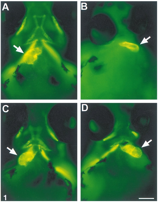
Fig. 1 Zebrafish mutants with defective notochords have randomized cardiac orientation. Ventral view of embryos stained with MF20 (Gonzalez-Sanchez and Bader, 1984) to view the cardiac ventricle and outflow tract, indicated by arrow. Atrium is lightly stained and out of the focal plane. Anterior is at the top. (A) Phenotypically wild-type embryo from no tail cross, displaying normal cardiac left–right orientation. (B) Floating head (flh) mutant with cardiac left –right reversal and smaller area of staining. (C) ntl mutant with a normal heart. (D) ntl mutant with a left–right reversed heart. Scale bar represents 100 μm.
Reprinted from Developmental Biology, 177, Danos, M.C. and Yost, H.J., Role of notochord in specification of cardiac left-right orientation in zebrafish and Xenopus, 96-103, Copyright (1996) with permission from Elsevier. Full text @ Dev. Biol.