
IMAGE
Fig. S2
- ID
- ZDB-IMAGE-090513-17
- Publication
- Trapani et al., 2009 - Synaptojanin1 is required for temporal fidelity of synaptic transmission in hair cells
- All Figures
- Figures for Trapani et al., 2009
Image
|
Figure Caption
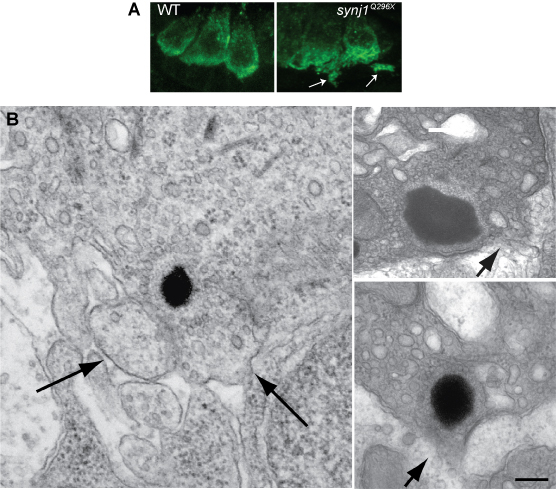
Fig. S2 Examples of loss of integrity of the plasma membrane near mutant synj1Q296X ribbons. (A) Confocal optical sections of inner ear hair cells labeled with anti-Vglut3 antibody in wild type and synj1Q296X larvae. (B) Left panel, blebbing seen in an synj1Q296X inner ear hair cell at the TEM level. Right panels, two ribbons in synj1Q296X neuromast hair cells. Arrows indicate membrane protrusions or areas that appear to the disintegrating. Scale bar, 125 nm in left panel; 100 nm in top right panel; 200 nm in lower right panel.
Acknowledgments
This image is the copyrighted work of the attributed author or publisher, and
ZFIN has permission only to display this image to its users.
Additional permissions should be obtained from the applicable author or publisher of the image.
Full text @ PLoS Genet.