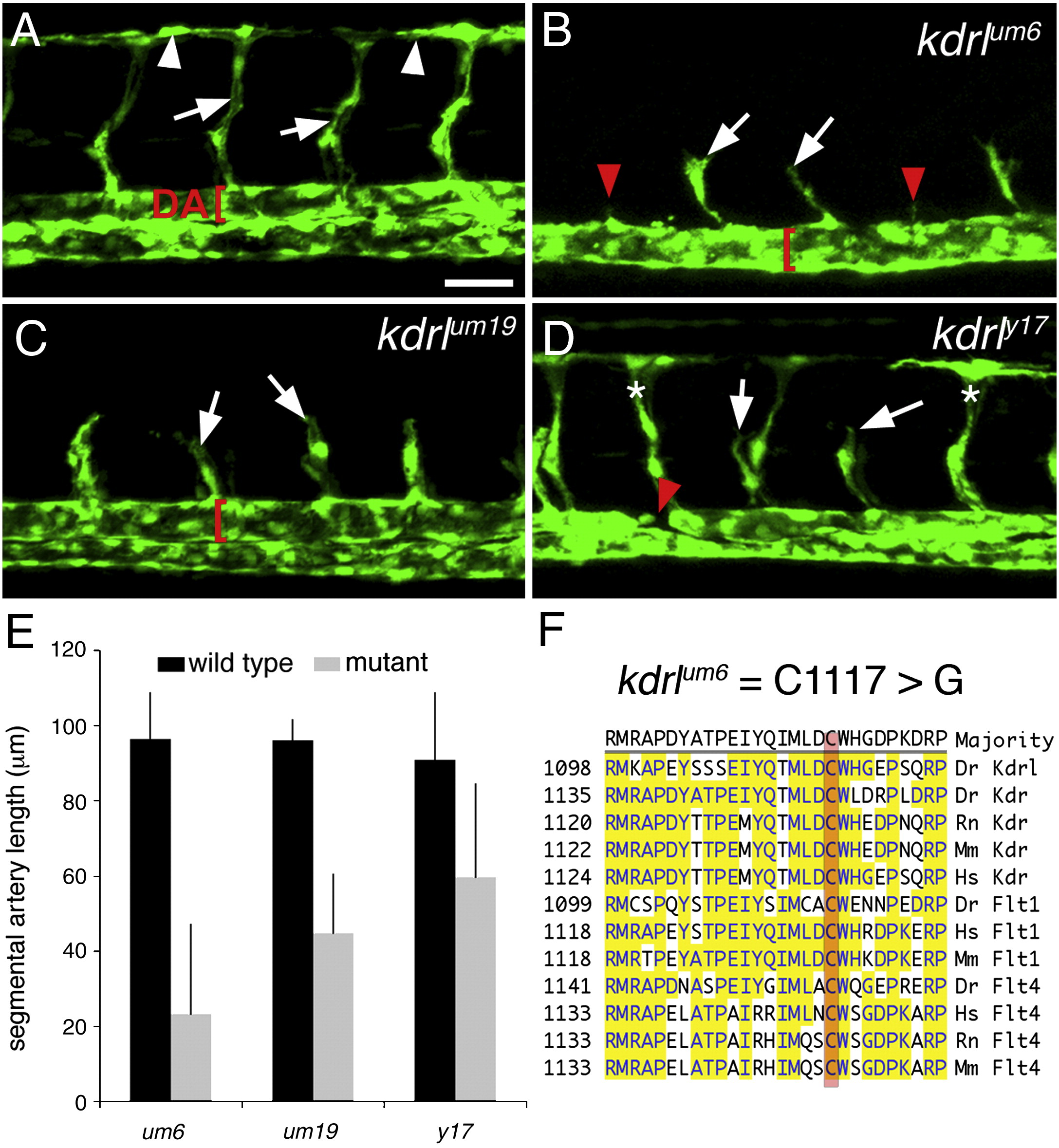
Fig. 2 Mutations in kdrl affect segmental artery formation. (A–D) Confocal fluorescent micrographs of zebrafish embryonic trunk blood vessels at 30 h post fertilization. Lateral views, anterior to the left, dorsal is up. Scale bar is 50 μM. (A) Wild type Tg(fli1a:egfp)y1 diploid sibling embryo. Arrowheads indicate DLAV. Arrows denote segmental arteries and dorsal aorta (DA) is indicated by a red bracket. (B) Segmental arteries in a kdrlum6;Tg(fli1a:egfp)y1 diploid mutant embryo. Partial segmental arteries indicated by white arrows. Red arrowheads indicate somite boundaries at which segmental arteries failed to sprout. Red bracket indicates a trunk vessel with a single lumen. (C) Partial segmental artery formation (white arrows) in a kdrlum19;Tg(fli1a:egfp)y1 diploid mutant embryo. A well-formed dorsal aorta (red bracket) is apparent in this embryo. (D) A kdrly17;Tg(fli1a:egfp)y1 diploid mutant embryo with both fully formed (indicated by asterisks) and partial segmental arteries (white arrows). The red arrow indicates region of the dorsal aorta that is discontinuous. (E) Quantification of sprout length in kdrl mutant embryos and their wild type siblings. (F) Location of the um6 mutation within the zebrafish Kdrl cytoplasmic domain. ClustalW alignment of conserved regions from Vegf-receptor-2/Kdr and Vegf-receptor-3/Flt4 from zebrafish (Dr), human (Hs), rat (Rn), and mouse.
Reprinted from Developmental Biology, 329(2), Covassin, L.D., Siekmann, A.F., Kacergis, M.C., Laver, E., Moore, J.C., Villefranc, J.A., Weinstein, B.M., and Lawson, N.D., A genetic screen for vascular mutants in zebrafish reveals dynamic roles for Vegf/Plcg1 signaling during artery development, 212-226, Copyright (2009) with permission from Elsevier. Full text @ Dev. Biol.

