
|
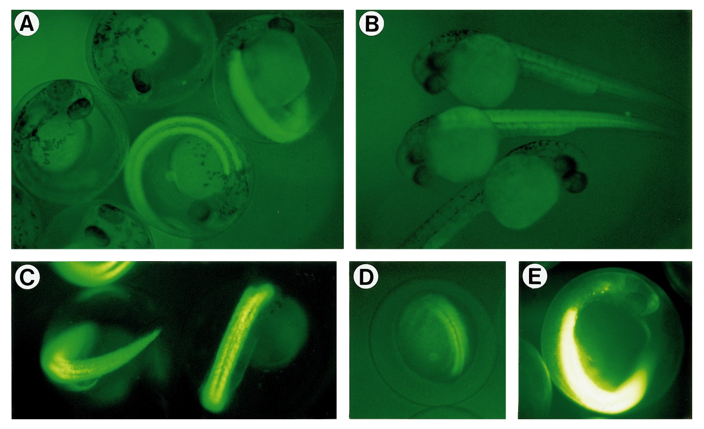
Fig. 4 Expression of GFP in transgenic embryos generated using α-actin–GFP. Embryos were viewed through their chorions, except for (B) where chorions were removed. (A) 30-h-old embryos of a B-rank transgenic line. Nonfluorescent embryos are nontransgenic siblings. (B) 28-h-old embryos of C-rank and D-rank transgenic lines. A nontransgenic embryo is also shown. (C) 26-h-old embryos of an A-rank transgenic line. (D) A 13-h-old embryo of an A-rank transgenic line. (E) A 26-h-old embryo of the most fluorescent line (A-rank). Film-exposure time for this figure was much shorter than that in other figures. Due to the high fluorescence of the muscles, other regions of the embryo are clearly visible.
Reprinted from Developmental Biology, 192, Higashijima, S., Okamoto, H., Ueno, N., Hotta, Y., and Eguchi, G., High-frequency generation of transgenic zebrafish which reliably express GFP in whole muscles or the whole body by using promoters of zebrafish origin, 289-299, Copyright (1997) with permission from Elsevier. Full text @ Dev. Biol.