
Fig. 4
- ID
- ZDB-IMAGE-090504-22
- Publication
- Mich et al., 2009 - Germ cell migration in zebrafish is cyclopamine-sensitive but Smoothened-independent
- All Figures
- Figures for Mich et al., 2009
|
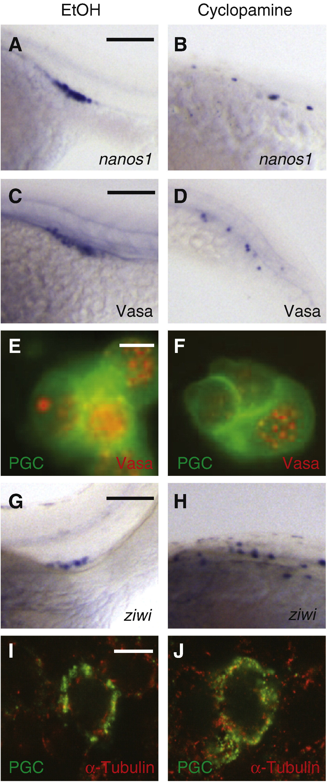
Fig. 4 Cyclopamine does not disrupt the subcellular organization or maturation of PGCs. (A, B) PGC-specific expression of nanos1, a maternally derived determinant of PGC specification, in ethanol- or cyclopamine-treated embryos. (C, D) Expression of Vasa protein, which is translated specifically in PGCs after 4 hpf. (E, F) Perinuclear granules containing Vasa-dsRedex protein (red) in EGFP-expressing PGCs (green). (G, H) PGC-specific expression of ziwi, which is transcribed in PGCs between 24 and 48 hpf. (I, J) Confocal immunofluorescence imaging of α-tubulin (red) and farnesylated EGFP (green) in the PGCs of Tg(kop:EGFP-F-nanos1–3′UTR) embryos. Developmental stages: A–F, 24 hpf; G–H, 48 hpf; I–J, 6 hpf. Scale bars: A–D and G, H, 100 μm; E–F, 20 μm; I, J, 10 μm.
Reprinted from Developmental Biology, 328(2), Mich, J.K., Blaser, H., Thomas, N.A., Firestone, A.J., Yelon, D., Raz, E., and Chen, J.K., Germ cell migration in zebrafish is cyclopamine-sensitive but Smoothened-independent, 342-354, Copyright (2009) with permission from Elsevier. Full text @ Dev. Biol.