
Fig. 1
- ID
- ZDB-IMAGE-090504-19
- Publication
- Mich et al., 2009 - Germ cell migration in zebrafish is cyclopamine-sensitive but Smoothened-independent
- All Figures
- Figures for Mich et al., 2009
|
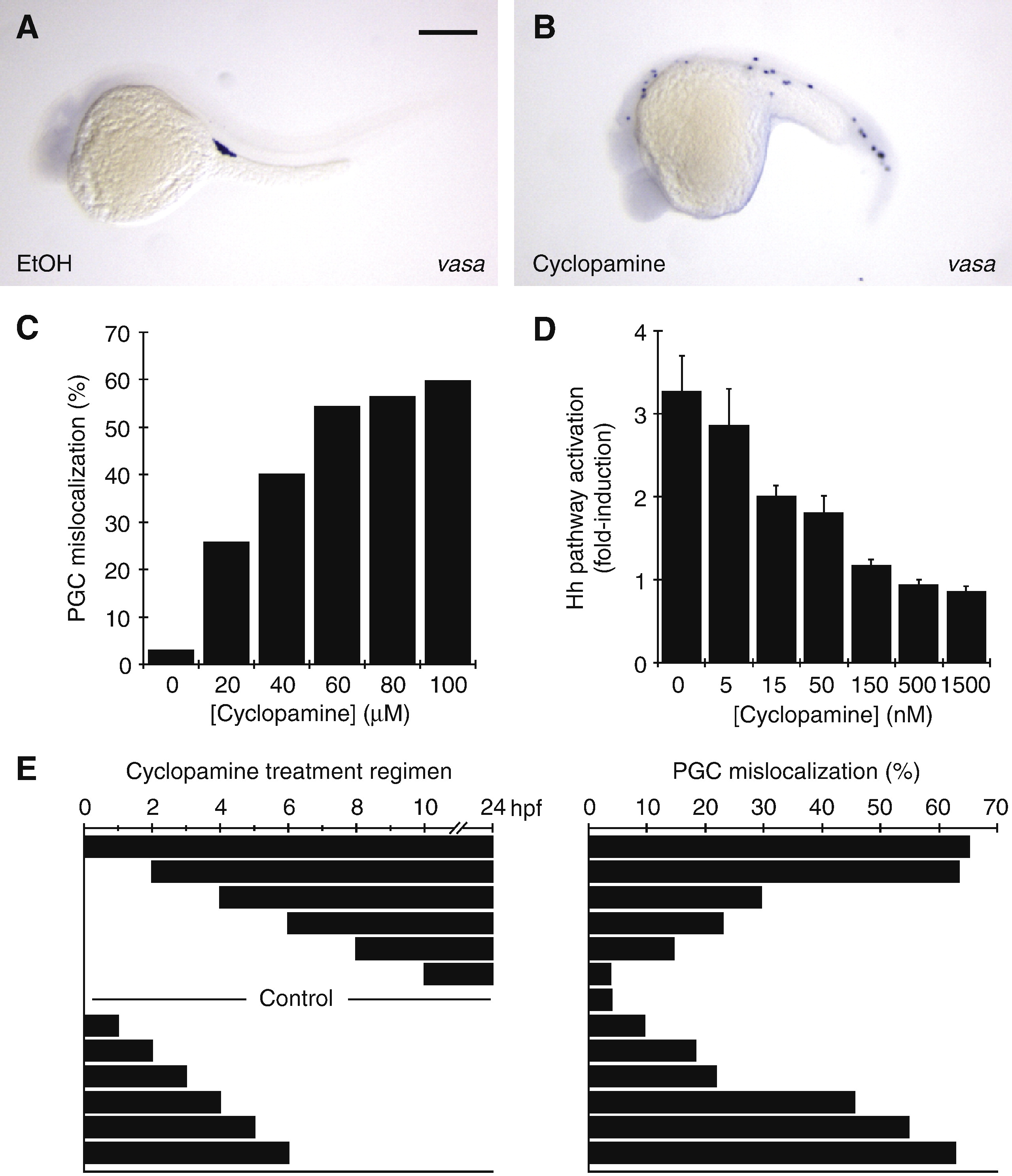
Fig. 1 Cyclopamine treatment induces zebrafish PGC mislocalization. (A, B) PGCs localize to the presumptive gonad in the ethanol-treated control embryos, as indicated by the distribution of vasa-expressing cells. In contrast, PGCs are widely dispersed in embryos treated with 60 μM cyclopamine. Cyclopamine and ethanol treatments were initiated at the one-cell stage and the resulting phenotypes at 24 hpf are shown. Scale bar: 200 μm. (C) Dose–response profile for cyclopamine-induced PGC mislocalization. At least 400 PGCs were scored at 24 hpf for each cyclopamine concentration. (D) Dose–response profile for cyclopamine inhibition of Shh signaling in Smo-/- mouse embryonic fibroblasts transfected with zebrafish smo. Data are the average of triplicate samples, with error bars representing the standard deviations. (E) Temporal window of cyclopamine action with respect to PGC mislocalization, as determined by a survey of treatment regimens. At least 500 PGCs were scored at 24 hpf for each time course of cyclopamine exposure.
Reprinted from Developmental Biology, 328(2), Mich, J.K., Blaser, H., Thomas, N.A., Firestone, A.J., Yelon, D., Raz, E., and Chen, J.K., Germ cell migration in zebrafish is cyclopamine-sensitive but Smoothened-independent, 342-354, Copyright (2009) with permission from Elsevier. Full text @ Dev. Biol.