
Fig. S10
- ID
- ZDB-IMAGE-090501-70
- Publication
- de Pater et al., 2009 - Distinct phases of cardiomyocyte differentiation regulate growth of the zebrafish heart
- All Figures
- Figures for de Pater et al., 2009
|
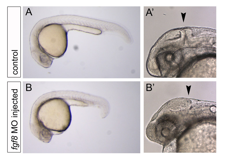
Fig. S10 fgf8 MO injection results in reduced Fgf signaling. (A,A′,B,B′) Live images of control embryos (A,A′) or fgf8 MO-injected embryos (B,B′). As previously shown by Draper et al. (Draper et al., 2001), the fgf8 MO used generates a range of phenotypic classes (Draper et al., 2001). In our experiments, injected embryos exhibited phenotypes similar to those described as class 1-3 phenotypes, most typically the class 2 phenotype (Draper et al., 2001). fgf8 MO-injected embryos were selected for the absence of the midbrain-hindbrain boundary, indicated by the black arrowhead. Approximately 40% of the injected embryos presented this phenotype and only these were imaged in the developmental timing assay.