
|
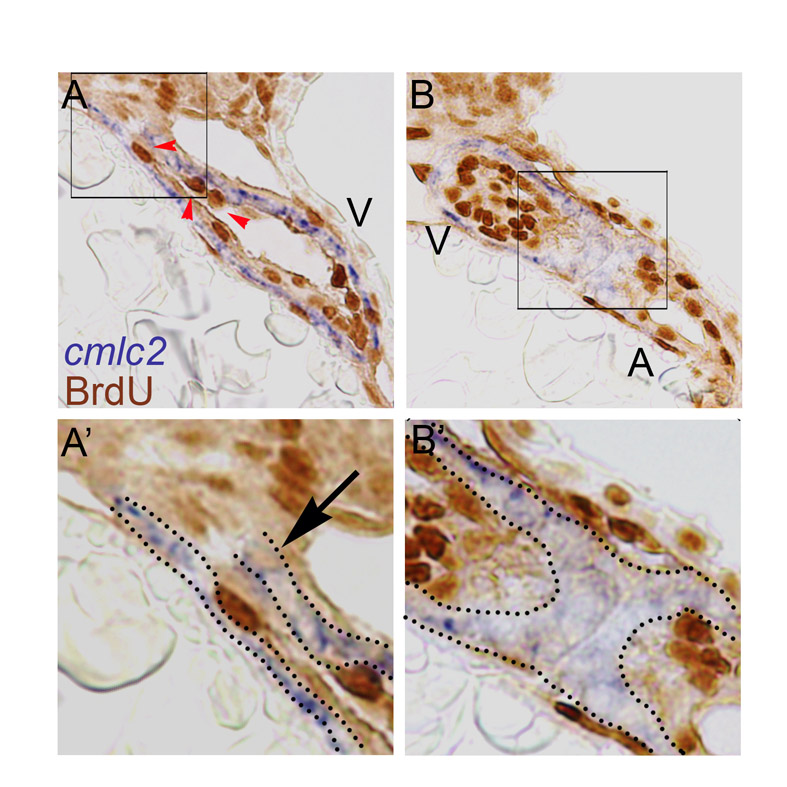
Fig. S2 Double labeling of BrdU and cmlc2. (A,A′,B,B′) Representative sections of BrdU-soaked embryos from 24 hpf until 48 hpf stained for cmlc2 in blue and BrdU in brown. These sections show that in the tissue expressing the cardiac marker cmlc2 very few cells are positive for BrdU (black arrow). The tissue lining the myocardium (the endocardium) is negative for cmlc2 and is highly proliferative, indicated by red arrowheads. Quantitation of the number of BrdU-positive cells within the cmlc2-positive area resulted in a similar number to that previously described in the results section (6±2, mean±s.e.m., n_5).