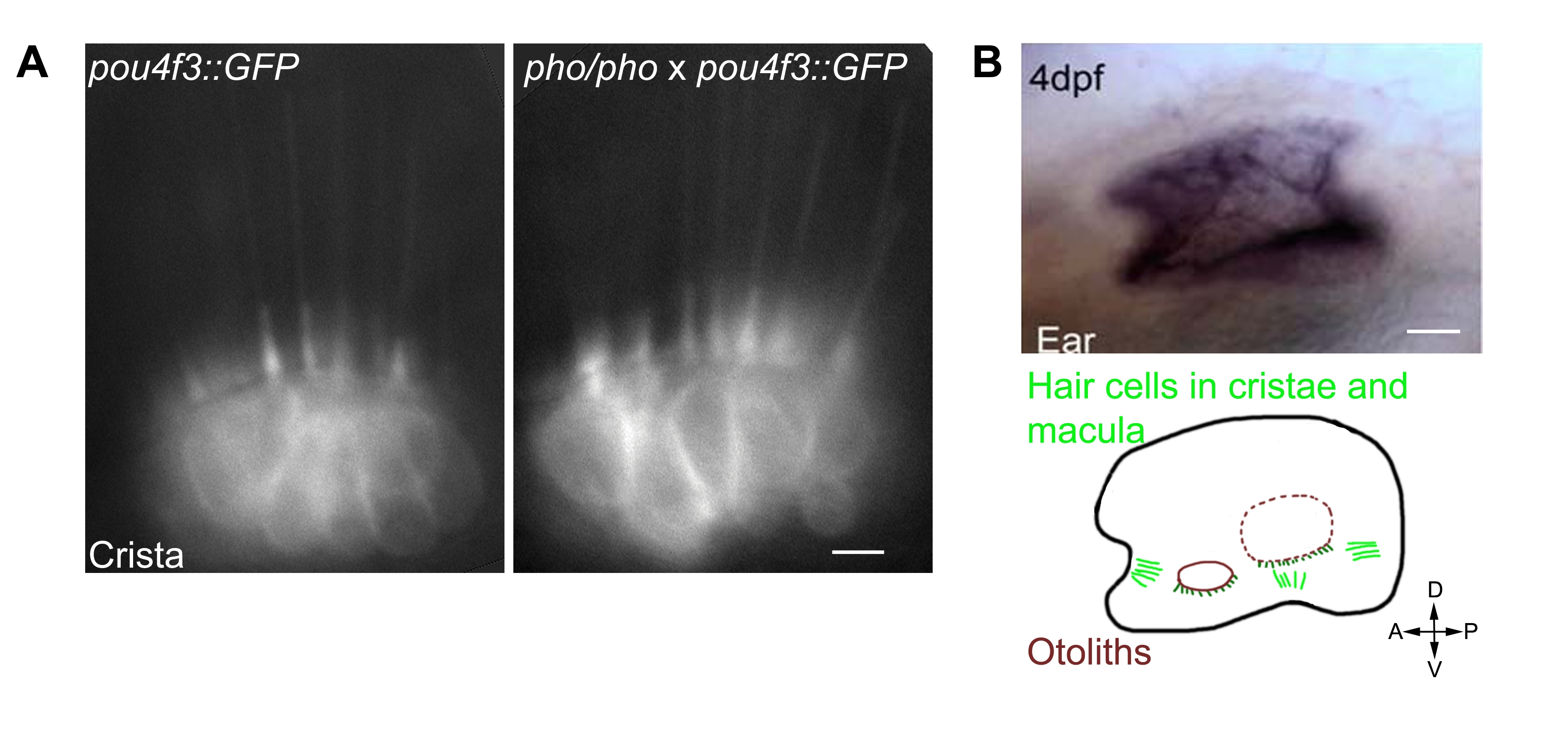
Fig. S2 Hair cells seem unaffected in the phoenix mutant ear. Wild-type expression of the phoenix mRNA and protein. A. Live images in pou4f3;;GFP transgenic animals of hair cells in a crista of a wild-type (left panel) and a phoenix mutant (right panel) ear. B. In situ hybridization with an antisense probe against phoenix in the inner ear of a 4dpf old embryo (top panel). A camera lucida drawing (lower panel) shows the positions of the two otoliths (red), of the cristae's (light green) and the maculae's (dark green) hair cells. The AP/DV ventral orientation is indicated. C. Immunofluorescence on cultured zebrafish cells (Pac2) using EX1 (green in the top and bottom left panels) or EX7 (green in the top and bottom right panels) antibodies. To highlight the ER, we co-stained with an antibody against PDI (ER-associated protein disulfide isomerase, red in middle and bottom left and right panels). DAPI was used to counterstain nuclei (blue in the two bottom left and right panels). Merged images (bottom, left and right panels) showed a cytoplasmic staining of phoenix, which was more concentrated in the perinuclear region. It appeared excluded from the ER, as there was no co-localization with DPI. Note how phoenix protein was upregulated in cells preparing to enter division (white arrow in the top right panel). D. Western blot on PAC2 cell extracts with two rabbit polyclonal antibodies, raised against an epitope encoded by exon 1 (EX1) or by exon 7 (EX7). A common band in the range of 95KDa was found with both antibodies (black arrow). - 10 microns in A, 50 microns in B and 3 microns in C.
Image
Figure Caption
Figure Data
Acknowledgments
This image is the copyrighted work of the attributed author or publisher, and
ZFIN has permission only to display this image to its users.
Additional permissions should be obtained from the applicable author or publisher of the image.
Full text @ PLoS Genet.

