
|
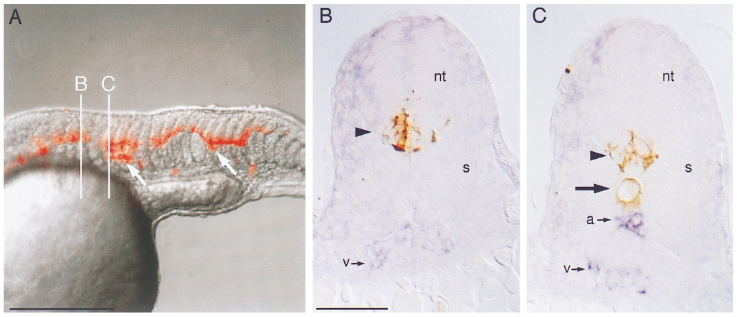
Fig. 8 Mosaic analysis of flh embryos transplanted with wild-type cells. (A) Composite rhodamine epifluorescence (red) and Nomarski (gray) videomicrograph of the trunk of a 25 somite flh mutant embryo that was transplanted at the blastoderm stage. In this embryo wildtype cells contributed primarily to neural tube and notochord (arrows). B and C indicate planes of cross-sectioning in panels (B) and (C). (B) Cross section of flh embryo in plane B. Transplanted wild-type cells labeled with biotin– peroxidase (brown, arrowhead) contributed to ventral neural tube (nt). A single population of flk-expressing cells (blue, v) resides ventrally. The somites (s) are fused beneath the neural tube. (C) Cross section of flh embryo in plane C. Transplanted wild-type cells contributed both to ventral neural tube (arrowhead) and to notochord (arrow). There are two populations of flk-expressing cells, a dorsal one (a) just beneath the notochord and a ventral one (v). Somites (s) are not fused. Dorsal is up, anterior is to the left in (A). Bars, 250 μm (A), 50 μm (B, C).
Reprinted from Developmental Biology, 183(1), Fouquet, B., Weinstein, B.M., Serluca, F.C., and Fishman, M.C., Vessel patterning in the embryo of the zebrafish: guidance by notochord, 37-48, Copyright (1997) with permission from Elsevier. Full text @ Dev. Biol.