
|
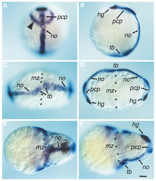
Fig. 5 Chordamesoderm in janus-mutant embryos at the end of gastrulation. (A,B) Wild-type. The neuroectodermal marker pax[zf-b] (arrowhead) (Krauss et al., 1991) allows to discriminate between ntl and hlx-1 expression domains. (C,D) janus-mutant embryo in which the shield contributes to the two blastoderms. Each shield results in the formation of a complete anterior axis. (E, F) janus-mutant embryo in which the shield is localized to one of the two blastoderms. Only the dorsal blastoderm forms a complete anterior axis. The ventral blastoderm lacks chordamesoderm. (A, C, E) Dorsal view. (B, D, F) Lateral view. Animal pole of the yolk cell is to the top. Hg, hatching gland expression domain of hgg-1; mz, marginal zone; no, notochord expression domain of ntl; pcp, prechordal plate expression domain of hlx-1; tb, tailbud expression domain of ntl. Black dots indicate the marginal zone between the two blastoderms. Scale bar is 100 μm.
Reprinted from Developmental Biology, 184(1), Abdelilah, S. and Driever, W., Pattern formation in janus-mutant zebrafish embryos, 70-84, Copyright (1997) with permission from Elsevier. Full text @ Dev. Biol.