
Fig. S1
- ID
- ZDB-IMAGE-090401-8
- Publication
- Sonawane et al., 2009 - Lgl2 and E-cadherin act antagonistically to regulate hemidesmosome formation during epidermal development in zebrafish
- All Figures
- Figures for Sonawane et al., 2009
|
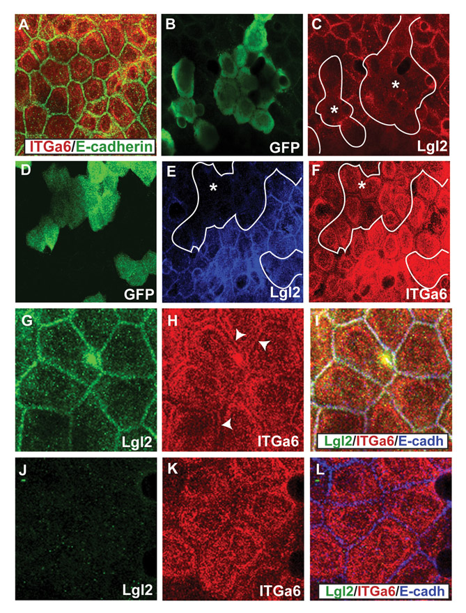
Fig. S1 Early localisation of Itga6 in lgl2 mutants and lgl2 morphant clones. (A-F) Co-immunostaining using anti-Itga6 (red) and anti-E-cadherin (A) antibodies; anti-GFP (B) and anti-Lgl2 (C) antibodies; anti-GFP (D), anti-Lgl2 (E) and anti-Itga6 (F) antibodies. The localisation of Itga6 is not altered in the basal epidermis of lgl2 larvae at 3 dpf (A). In the basal epidermal clones marked with GFP (B), lgl2 morpholino specifically knockdowns Lgl2 expression (C). In GFP-marked basal epidermal clones (D), wherein Lgl2 expression is attenuated (E), Itga6 localisation at the basal and lateral domains remains unperturbed. (G-L) Co-immunostainings using anti-Lgl2 (G,J), anti-Itga6 (H,K) and anti-E-cadherin (blue in the overlays; I,L) antibodies. In comparison to wild type (G-I), in the lgl2 mutant (J-L) at 3.75 dpf lateral Itga6 localisation is selectively altered. Asterisks, epidermal clones. Arrowheads in H mark the lateral Itga6 localisation in wild-type basal epidermis.