
|
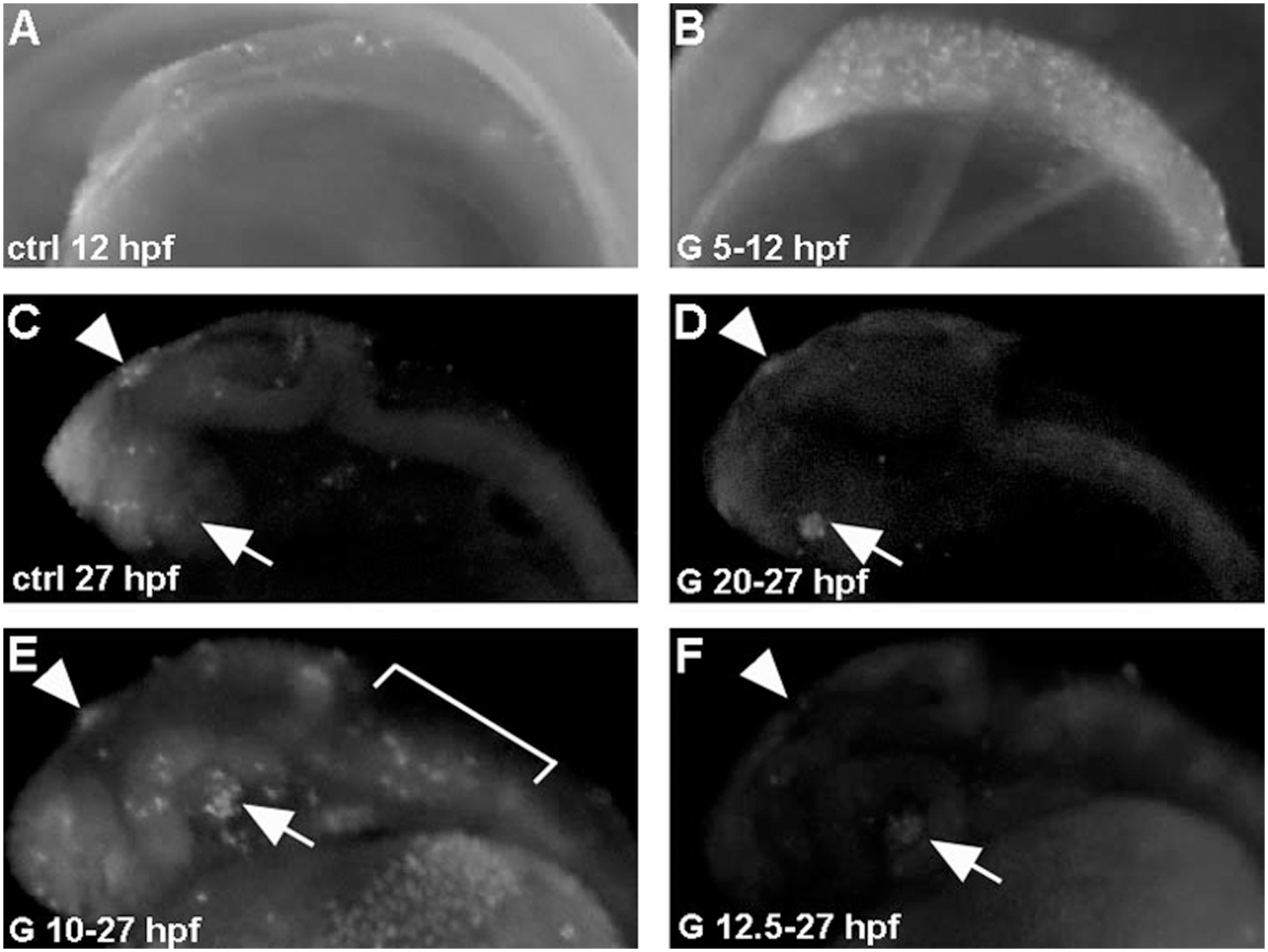
Fig. 2 Genistein-induced apoptosis is developmental stage-dependent.
Embryos were treated with 2.5 μM genistein for 7 hours starting at 5 hpf (B) or 20 hpf (D) and stained with acridine orange. A high number of apoptotic cells, compare with control (A), are detected in embryos treated from 5 hpf onwards. A similar localization (retina and epiphysis, arrow and arrowhead, respectively) and small number of apoptotic cells are detected in control (C) and genistein-treated embryos from 20 hpf onwards (D). (E) Treatment from 10 hpf onwards induces a strong apoptosis detected at 27 hpf (arrows), while a similar treatment started at 12.5 hpf did not induce apoptosis at 27 hpf (F).