
|
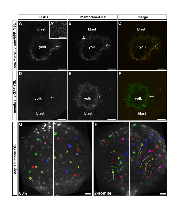
Fig. S3 Overexpression of Oep within the YSL does not rescue defective iYSN convergence movements in MZoep mutant embryos. (A-C) Oep-FLAG antibody staining in MZoep embryos overexpressing oep in the YSL during gastrulation. The YSL of MZoep embryos were injected with 400 pg oep-FLAG and 100 pg membrane-bound gap43-GFP. Confocal transversal sections showing expression of Oep-FLAG within the YSL (A), which colocalize with membrane-bound GFP (B,C; arrows). (A′) Oep-FLAG antibody staining in an MZoep embryo injected with 200 pg oep-FLAG at the one-cell stage, showing expression of Oep on the cell membranes of blastoderm cells. (D,E) MZoep embryo injected with 100 pg membrane bound-GFP into the YSL. Confocal transversal section showing no detection of the FLAG tag (D), whereas membrane-bound GFP is strongly expressed in the YSL (E,F, arrows). (A,D) Flag antibody staining. (B,E) Membrane-bound GFP. (C,F) Merged image of FLAG and GFP channels. (G,H) iYSN trajectories in MZoep mutant injected with oep-FLAG into the YSL at 80% epiboly (8 hpf; G, startpoint of tracks) and two-somite stage (11 hpf; H, endpoint of tracks). Images are z-projections. Some nuclear trajectories obtained using Motion Tracking Software are shown. Circles indicate the endpoint of each track. White line marks the dorsal midline of the embryo. Animal is towards the top. Blast, blastoderm. Scale bars 40 μm in A-F; 50 μm in G,H.