
|
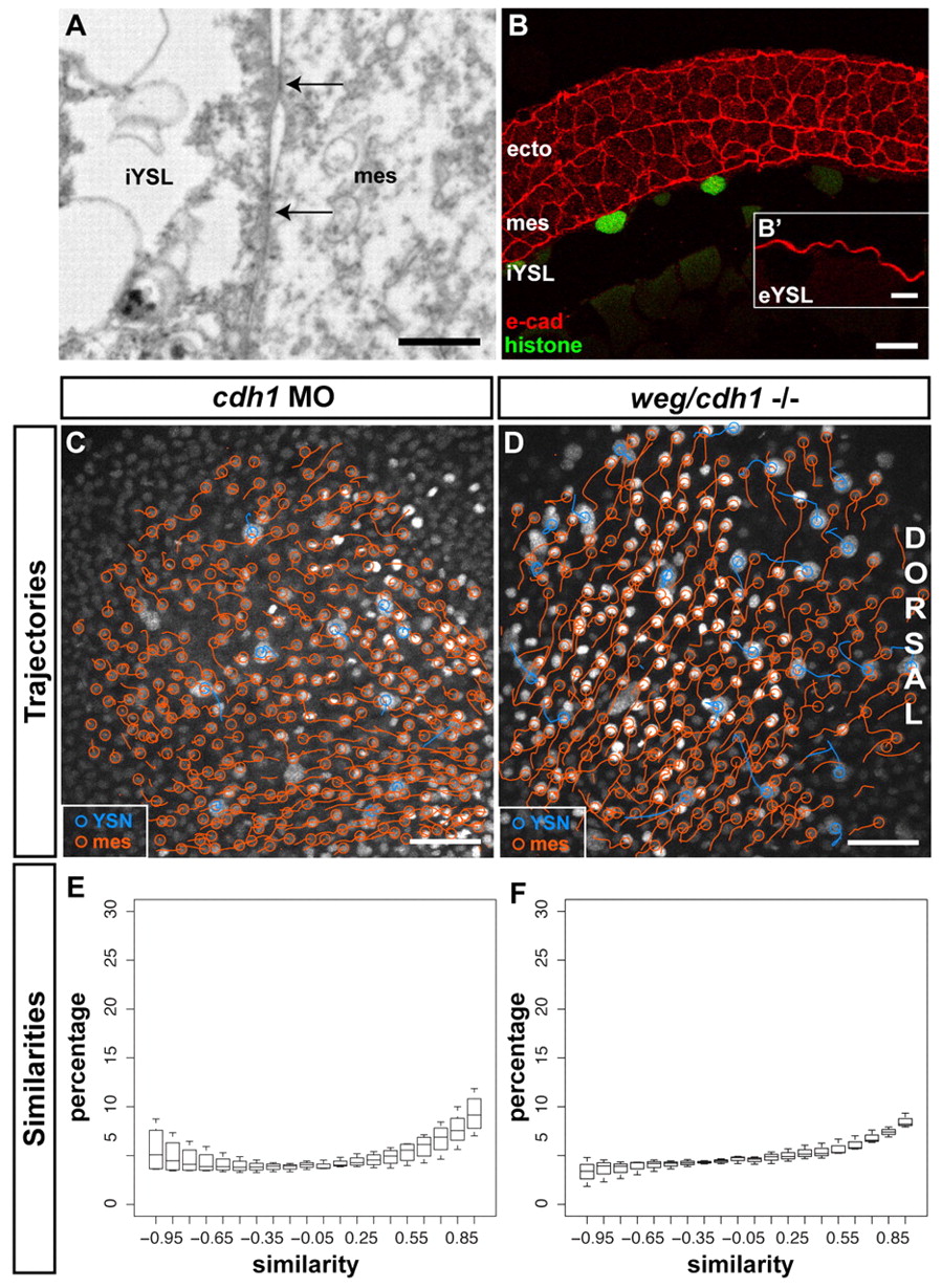
Fig. 5 E-cadherin is required for convergence movement coordination between iYSN and mesendoderm. (A) Sagittal section obtained by transmission electron microscopy of a wild-type embryo at 80% epiboly stage (8 hpf) showing a mesendoderm progenitor in close contact with the iYSL plasma membrane. Note the presence of electron-dense regions (arrows) of tight contact between the mesendoderm progenitor and the iYSL plasma membrane. Animal pole is towards the right. (B) E-cadherin antibody staining of transversal (B) and sagittal (B′) sections of wild-type gastrulating embryos. E-cadherin is localized at ectoderm, mesendoderm and YSL plasma membranes. (B′) Magnification of the eYSL region. (C,D) Trajectories of iYSN (blue tracks) and mesendoderm progenitors (orange tracks) in e-cadherin morphant (C) and weg/e-cadherin mutant (D) embryos at mid-gastrulation stages (7-8 hpf). Images are z-projections. Dorsal is towards the right. (E,F) Quantification of the similarity between iYSN and mesendoderm convergence movements in e-cadherin morphants (E) and weg/e-cadherin mutants (F) at mid-gastrulation stages (7-8 hpf). Histograms of the similarity values were generated separately for each embryo. Boxplots show the distribution of the bin heights among the embryos. (E) In e-cadherin morphants, 35% of the similarity values are higher than 0.5 (n=4 embryos). (F) In weg/e-cadherin mutants, 35% of the similarity values are higher than 0.5 (n=3 embryos). e-cad, e-cadherin; ecto, ectoderm progenitor; mes, mesendoderm progenitor. Scale bars: 2 μm in A; 20 μm in B; 10 μm in B′; 50 μm in C,D.