
Fig. 1
- ID
- ZDB-IMAGE-090325-6
- Publication
- Goessling et al., 2009 - Genetic interaction of PGE2 and Wnt signaling regulates developmental specification of stem cells and regeneration
- All Figures
- Figures for Goessling et al., 2009
|
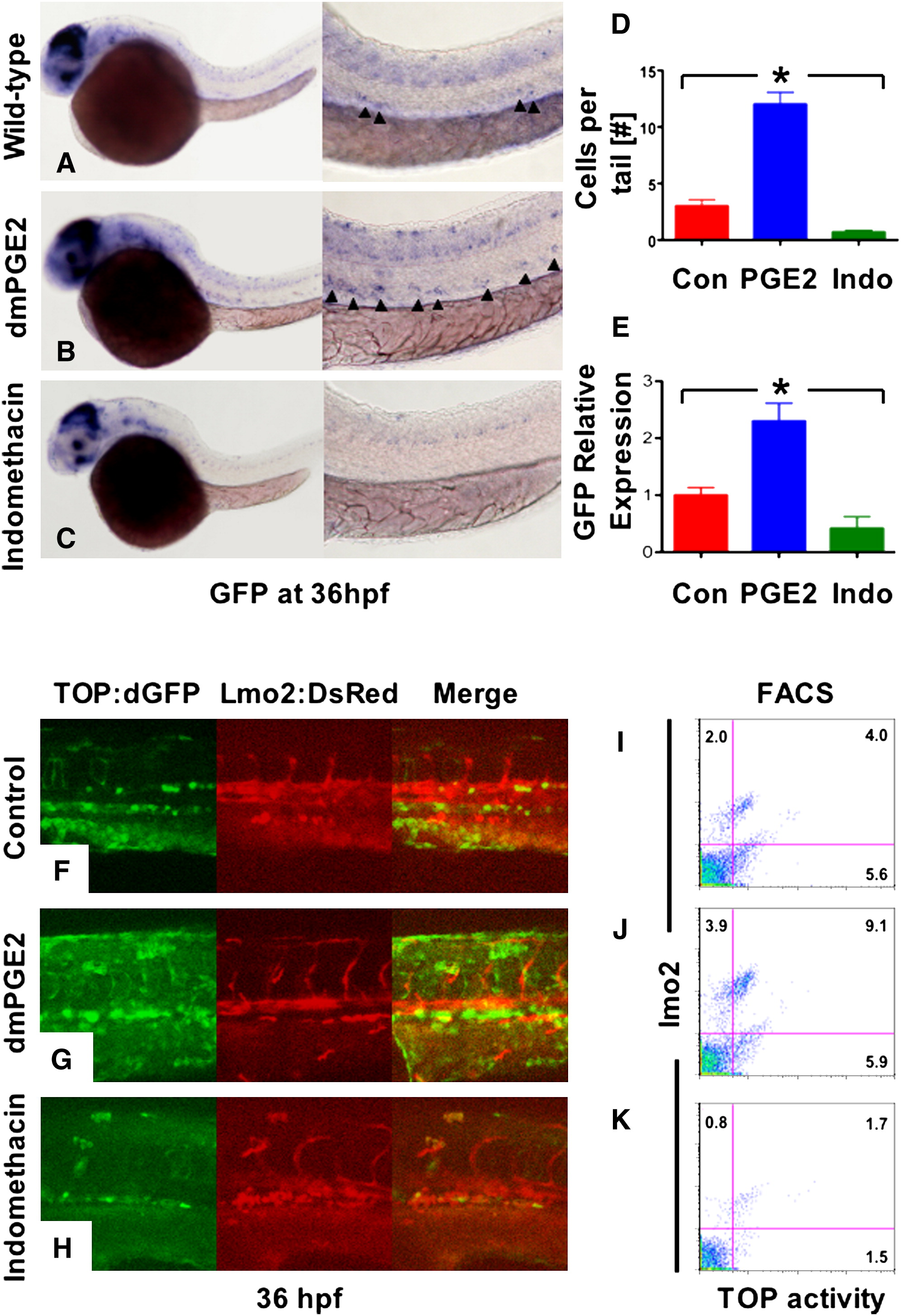
Fig. 1 Prostaglandin Levels Directly Affect wnt Activity in Zebrafish Embryos
(A–C) In situ hybridization for GFP in TOP:dGFP wnt reporter embryos at 36 hpf shows widespread wnt activity; inset, close-up of GFP+ (black arrowheads) cells in the AGM. 10 μM dmPGE2 enhanced GFP expression throughout the embryo, while 10 μM indo decreased global wnt activity, and in the AGM.
(D) Quantification (mean ± SD) of total GFP+ cells in the major trunk vessels and (E) qPCR analysis for GFP in whole embryo lysates following exposure to dmPGE2 or indo versus vehicle con reveals significant alterations in wnt activity (*significant (sig) across treatment groups, ANOVA, p < 0.05, n = 10/treatment).
(F–H) Representative confocal microscopy images of the AGM region of TOP:dGFP; lmo2:DsRed embryos following exposure to con, dmPGE2, or indo are shown; differences can be seen in the wnt-active (green, left column), HSC/endothelial (red, middle), and colocalized (merged, right) populations.
(I–K) Representative FL1 (green)/ FL2 (red) FACS plots of individual TOP:dGFP; lmo2:DsRed embryos after exposure to con, dmPGE2 or indo confirm the confocal analyses (summarized in Figure S2).
Reprinted from Cell, 136(6), Goessling, W., North, T.E., Loewer, S., Lord, A.M., Lee, S., Stoick-Cooper, C.L., Weidinger, G., Puder, M., Daley, G.Q., Moon, R.T., and Zon, L.I., Genetic interaction of PGE2 and Wnt signaling regulates developmental specification of stem cells and regeneration, 1136-1147, Copyright (2009) with permission from Elsevier. Full text @ Cell