
IMAGE
Fig. S2
- ID
- ZDB-IMAGE-090320-7
- Publication
- Navratilova et al., 2009 - Systematic human/zebrafish comparative identification of cis-regulatory activity around vertebrate developmental transcription factor genes
- All Figures
- Figures for Navratilova et al., 2009
Image
|
Figure Caption
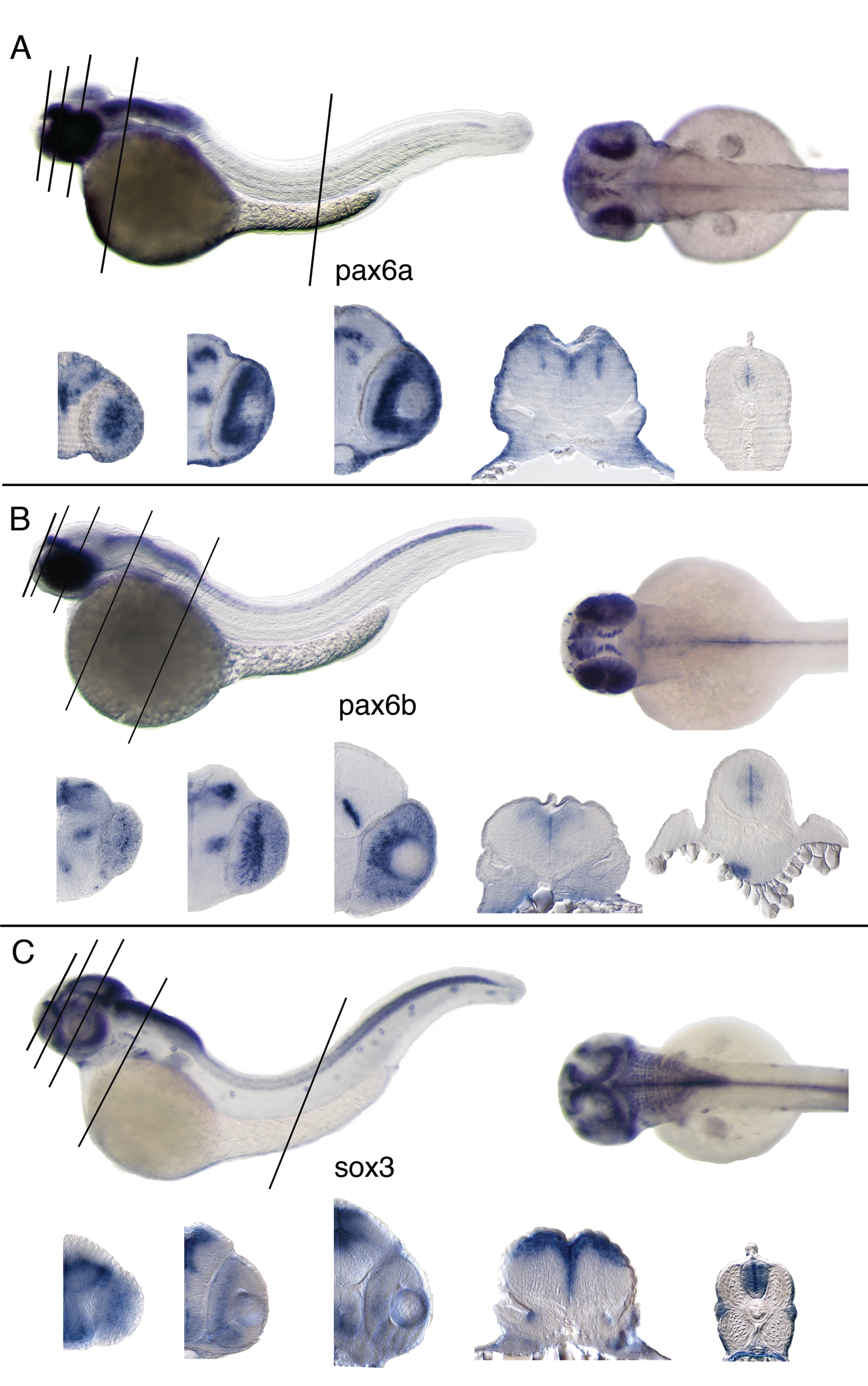
Fig. S2 Lateral, dorsal views and vibratome sections of whole mount RNA in situ hybridizations. 2 days old zebrafish embryos and 20 μm thick transversal vibratome sections below (levels indicated by black lines on the whole mount). A: pax6a; expressed in hindbrain, optic tectum, anterior spinal cord, retina, diencephalon. B: pax6b; detected in hindbrain, spinal cord, retina, diencephalon, pancreas. C: sox3; detected in CNS, particularly in ventricular zone of the neural tube also in retina, branchial arches, lateral line an inner ear.
Acknowledgments
This image is the copyrighted work of the attributed author or publisher, and
ZFIN has permission only to display this image to its users.
Additional permissions should be obtained from the applicable author or publisher of the image.
Reprinted from Developmental Biology, 327(2), Navratilova, P., Fredman, D., Hawkins, T.A., Turner, K., Lenhard, B., and Becker, T.S., Systematic human/zebrafish comparative identification of cis-regulatory activity around vertebrate developmental transcription factor genes, 526-540, Copyright (2009) with permission from Elsevier. Full text @ Dev. Biol.