
Fig. 5
- ID
- ZDB-IMAGE-090320-27
- Publication
- Stedman et al., 2009 - A functional interaction between Irx and Meis patterns the anterior hindbrain and activates krox20 expression in rhombomere 3
- All Figures
- Figures for Stedman et al., 2009
|
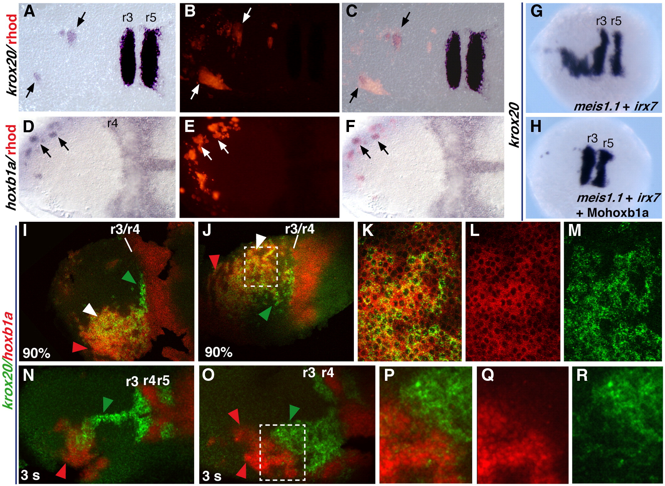
Fig. 5 Mechanisms of krox20 activation downstream of irx7 and meis1.1. (A–F) Host embryos processed for rhodamine (B, E) and krox20 (A) or hoxb1a (D) staining, after transplantation of cells form donor embryos injected with rhodamindextran, irx7 and meis1.1. Panels C and F are merged images of panels A and B, or panels C and D, respectively. (G, H) Embryos injected with irx7 and meis1.1 RNAs and with (H) or without (G) a morpholino for hoxb1a (Mohoxb1a). (I–R) Embryos coinjected with irx7 and meis1.1 RNAs and processed for fluorescent ISH. K–M and P–R are higher magnifications of the region squared in J and O, respectively, showing krox20 staining (green, M,R), hoxb1a staining (red, L, Q) or both (merge, K, P). All pictures are dorsal views, with anterior to the left, of flat-mounted (A–F and I–R) or whole (G, H) embryos. Probes are indicated on the left and colour-coded. All embryos are at the 5 s stage, except when otherwise stated at the bottom left of the picture. In panels I–R, green, red and white arrowheads point to cells showing ectopic expression of krox20 only, hoxb1a only or both, respectively.
Reprinted from Developmental Biology, 327(2), Stedman, A., Lecaudey, V., Havis, E., Anselme, I., Wassef, M., Gilardi-Hebenstreit, P., and Schneider-Maunoury, S., A functional interaction between Irx and Meis patterns the anterior hindbrain and activates krox20 expression in rhombomere 3, 566-577, Copyright (2009) with permission from Elsevier. Full text @ Dev. Biol.