
|
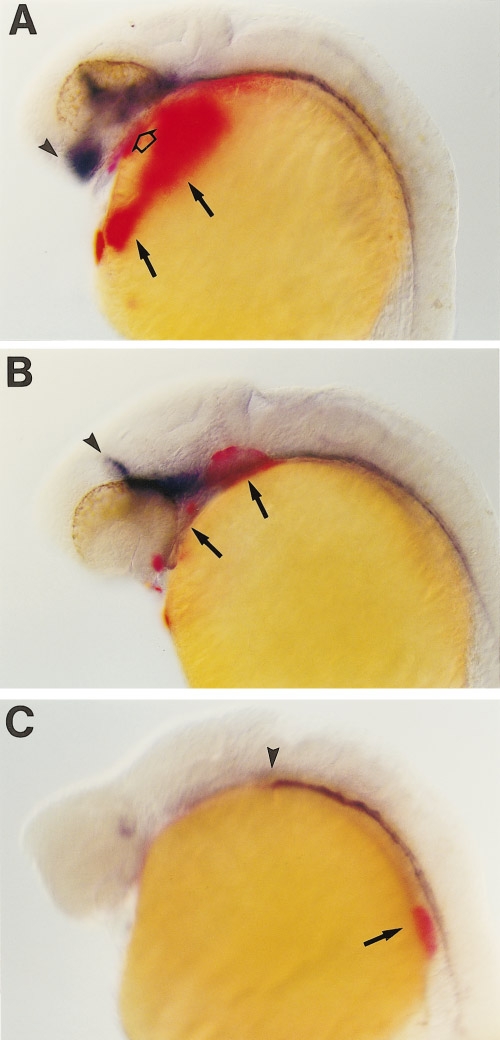
Fig. 9 Cyclopic embryos lack correctly positioned hatching glands. Ethanol-treated embryos at the 24-h stage were simultaneously stained for hgg-1 (red) and shh (blue) expression; hgg1 is specifically expressed in hatching glands. (A) Ethanol-treated embryo with narrowly spaced eyes have hatching glands over the yolk (arrows). Some hgg1-expressing cells were incorrectly positioned under the forebrain (open arrow). (B) Cyclopic embryo which lacks the wild-type pattern of hgg-1 expression on the yolk; hatching glands are instead found at the level of the forebrain and midbrain (arrows). Note that the anterior-most expression of shh in the hypothalamus is missing in this embryo (compare arrowheads marking anterior limit of shh expression in A and B). (C) Profoundly cyclopic embryo which lacks shh expression in the fore- and hgg-1-expressing cells are located in the trunk in this embryo (arrow). Orientation of embryos is anterior to the left and dorsal up. Arrowheads, anterior limits of shh expression.
Reprinted from Developmental Biology, 201, Blader, P. and Strähle, U., Ethanol impairs migration of the prechordal plate in the zebrafish embryo, 185-201, Copyright (1998) with permission from Elsevier. Full text @ Dev. Biol.