
Fig. S3
- ID
- ZDB-IMAGE-090224-26
- Publication
- Chan et al., 2009 - Functional analysis of the evolutionarily conserved Cis-regulatory elements on the Sox17 gene in zebrafish
- All Figures
- Figures for Chan et al., 2009
|
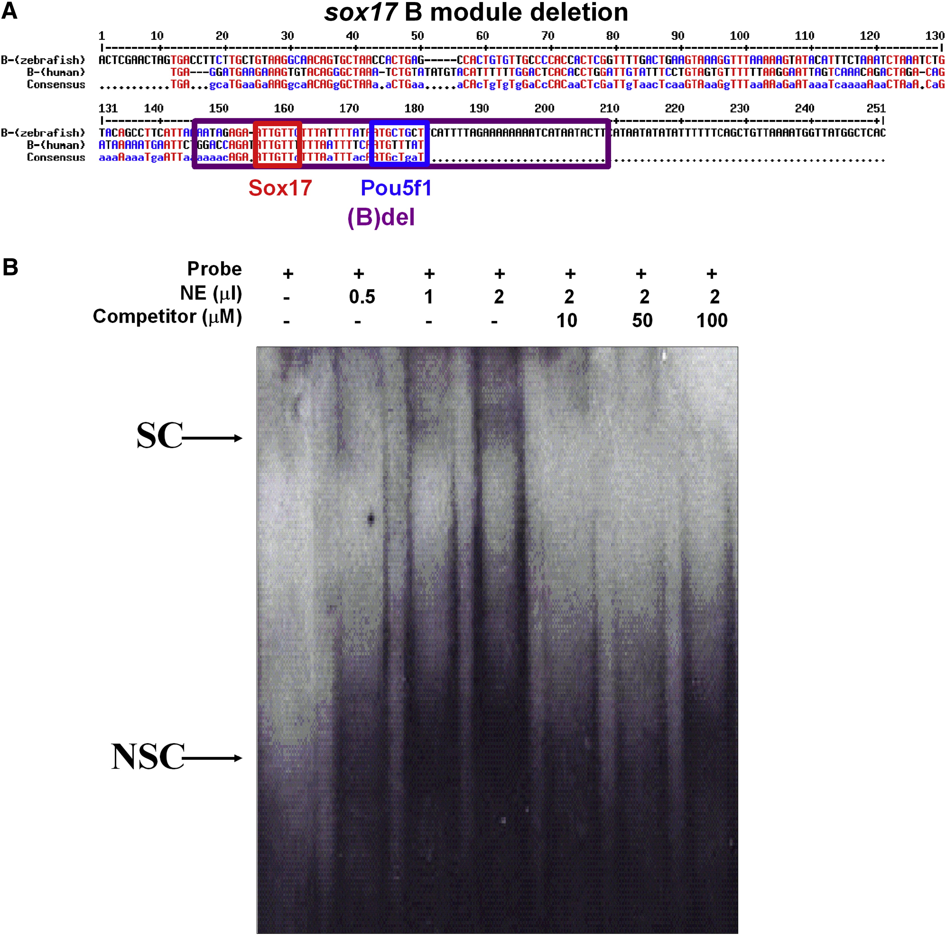
Fig. S3 Electrophoretic mobility shift assays of pou5f1 binding site on the sox17 cis-element module B. (A) The sequence of the B module, the red line indicated the probe for the binding assay. Red line indicated the sequence for making a probe. (B) A different amount of nuclear extract (0.5, 1 and 2 μl) was added into each reaction as indicated. A different amount of specific competitors (10, 50 and 100 μM) was added into the reaction as indicated. Two complexes were detected when the probe was added into nuclear extracts (NE). The upper band can be specifically competed with a specific competitor but not with a non-specific competitor that was denoted as “S”. The “NS” indicates the lower band, which is non-specific because it can be competed with both specific and non-specific competitors.
Reprinted from Developmental Biology, 326(2), Chan, T.M., Chao, C.H., Wang, H.D., Yu, Y.J., and Yuh, C.H., Functional analysis of the evolutionarily conserved Cis-regulatory elements on the Sox17 gene in zebrafish, 456-470, Copyright (2009) with permission from Elsevier. Full text @ Dev. Biol.