
IMAGE
Fig. S1
- ID
- ZDB-IMAGE-090220-9
- Publication
- Ota et al., 2009 - The roles of the FGF signal in zebrafish embryos analyzed using constitutive activation and dominant-negative suppression of different FGF receptors
- All Figures
- Figures for Ota et al., 2009
Image
|
Figure Caption
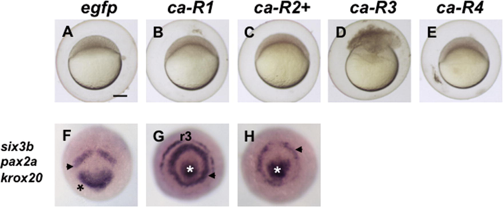
Fig. S1 Effects of ca-FGFRs at the blastula stage and later stages. (A–E) Dome-stage embryos injected with ca-FGFR mRNA. (F–H) Frontal views of embryos injected with egfp or ca-R mRNA (dorsal to the top), which were stained at the bud stage for six3b, pax2a, and krox20. Scale bar, 100 μm.
Acknowledgments
This image is the copyrighted work of the attributed author or publisher, and
ZFIN has permission only to display this image to its users.
Additional permissions should be obtained from the applicable author or publisher of the image.
Reprinted from Mechanisms of Development, 126(1-2), Ota, S., Tonou-Fujimori, N., and Yamasu, K., The roles of the FGF signal in zebrafish embryos analyzed using constitutive activation and dominant-negative suppression of different FGF receptors, 1-17, Copyright (2009) with permission from Elsevier. Full text @ Mech. Dev.