
Image
|
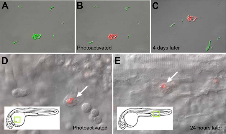
Figure Caption
Fig. S4 M. marinum expressing Kaede photoactivatable protein to mark and track infected macrophages in vivo. (A-B) Merged image of photoactivated (red) and non-photoactivated (green) bacteria in medium immediately before (A) and after (B) photoactivation. (C) Same bacteria still detectable four days later. (D) Single macrophage containing photoactivated M. marinum (red channel only) in yolk circulation valley of embryo, immediately after photoactivation. (E) The same bacteria detected 24 hours later in caudal vein. Insets show location in embryo.
Acknowledgments
This image is the copyrighted work of the attributed author or publisher, and
ZFIN has permission only to display this image to its users.
Additional permissions should be obtained from the applicable author or publisher of the image.
Reprinted from Cell, 136(1), Davis, J.M., and Ramakrishnan, L., The role of the granuloma in expansion and dissemination of early tuberculous infection, 37-49, Copyright (2009) with permission from Elsevier. Full text @ Cell