
|
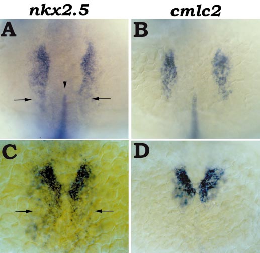
Fig. 2 cmlc2 is expressed in most, but not all, nkx2.5-expressing cells. (A, B) Dorsal views of embryos at the 14-somite stage, anterior at the top; in situ hybridization with nkx2.5 and no tail (A) or cmlc2 and no tail (B) riboprobes. no tail is expressed in the developing notochord (Schulte-Merker et al., 1994). (A) nkx2.5 expression in bilateral stripes of precardiac mesoderm extends slightly (arrows) beyond the anterior tip of the developing notochord (shown by no tail expression, arrowhead). This most posterior region has relatively weak expression of nkx2.5. (B) The posterior boundaries of the bilateral stripes of cmlc2 expression are aligned with the anterior tip of no tail expression. (C, D) Dorsal views of embryos at the 17-somite stage, anterior at the top; in situ hybridization with nkx2.5 (C) or cmlc2 (D) riboprobes. As the bilateral cardiac primordia bend toward each other, the most posterior nkx2.5-expressing cells (C, arrows) do not express cmlc2 (D). Again, these most posterior cells express relatively weak levels of nkx2.5 (C).
Reprinted from Developmental Biology, 214(1), Yelon, D., Horne, S.A., and Stainier, D.Y.R., Restricted expression of cardiac myosin genes reveals regulated aspects of heart tube assembly in zebrafish, 23-37, Copyright (1999) with permission from Elsevier. Full text @ Dev. Biol.